
Архив офтальмологии Украины Том 13, №2, 2025
Вернуться к номеру
Неврит зорового нерва як ускладнення запальної патології приносових порожни
Авторы: Євчев Ф.Д., Єпішева С.М., Дьячкова З.Є., Терещенко А.А.
Одеський національний медичний університет, м. Одеса, Україна
Рубрики: Офтальмология
Разделы: Справочник специалиста
Версия для печати
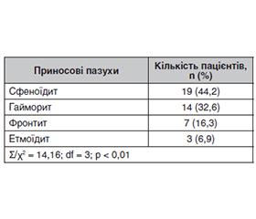
Актуальність. Орбітальні ускладнення синуситів — це група захворювань очної ямки, які розвиваються на тлі запальних уражень придаткових пазух носа. За даними багатьох авторів, перше місце посідає запалення верхньощелепних пазух, на другому місці — запалення решітчастих лабіринтів, на третьому — сфеноїдальних пазух. Мета: проаналізувати частоту виникнення і подати клінічну характеристику невриту зорового нерва, що виник унаслідок запалення приносових порожнин. Матеріали та методи. Обстежені 43 пацієнти (43 ока) — 14 жінок (32,6 %) і 29 чоловіків (67,4 %) віком від 19 до 58 років з діагнозом невриту зорового нерва. Після огляду та збору анамнезу ми запідозрили, що неврит виник унаслідок запалення приносових порожнин. У всіх пацієнтів спостерігався монолатеральний процес. Проводилися загальноклінічне обстеження, визначення гостроти зору, поля зору, офтальмоскопія, комп’ютерна томографія, передня і задня риноскопія, фарингоскопія, рентгеноскопія в прямих проєкціях і прицільній боковій проєкції, ендоскопія. Результати. Основні клінічні скарги пацієнтів на погіршення або втрату зору, головний біль, частіше в зоні скроні й потилиці та при нахилі голови вперед, ускладнення дихання і виділення з носа, слабкість, температуру дають нам підстави запідозрити запальний процес у приносових пазухах. Найбільш поширеною причиною виникнення невриту зорового нерва внаслідок запалення приносових пазух визнано сфеноїдити. Це пов’язано з тим, що найбільш часто зоровий нерв розташовується близько до задньої ґратчастої та клиноподібної пазух без контакту або поглиблення стінки. На другому місці — гайморити, на третьому — фронтити. Висновки. Сфеноїдит найбільш часто діагностується як причина серйозних ускладнень, як-от неврит зорового нерва і менінгіт. Запалення приносових пазух можуть давати дуже серйозні ускладнення: неврит зорового нерва, менінгіт, тромбоз печеристого синуса. У всіх випадках, коли це захворювання лише запідозрено, необхідно терміново вживати заходів щодо диференціальної діагностики й лікування. Найчастіше в запальний процес зорового нерва (неврит) залучена сфеноїдальна пазуха, що потребує негайного лікування для запобігання ускладненням.
Background. Orbital complications of sinusitis are a group of diseases of the eye socket that develop against the background of inflammatory lesions of the paranasal sinuses. According to many authors, inflammation of the maxillary sinuses ranks first followed by inflammation of the ethmoidal labyrinths and of the sphenoid sinuses. Objective: to analyze the frequency of occurrence and clinical characteristics of optic neuritis, which arose as a result of inflammation of the paranasal sinuses. Materials and methods. We examined 43 patients (43 eyes), 14 women (32.6 %) and 29 men (67.4 %) aged 19 to 58 years, with a diagnosis of optic neuritis. After examination and history taking, we suspected that neuritis was caused by inflammation of the paranasal sinuses. In all patients, a unilateral process was observed. A general clinical examination, determination of visual acuity, visual fields, ophthalmoscopy, computed tomography, anterior and posterior rhinoscopy, pharyngoscopy, radiography in direct projections and aiming lateral projection, endoscopy were carried out. Results. The main clinical complaints of patients about deterioration or loss of vision, headaches, more often in the area of the temple and the back of the head, and when tilting the head forward, difficulty breathing and discharge from the nose, weakness, temperature give us reason to suspect an inflammatory process in the paranasal sinuses. Sphenoiditis is recognized as the most common cause of optic neuritis due to inflammation of the paranasal sinuses. This is because the optic nerve is most often located close to the posterior ethmoid and sphenoid sinuses without contact or deepening of the wall. Sinusitis ranks second, followed by frontal sinusitis. Conclusions. Sphenoiditis is most often diagnosed as the cause of serious complications, such as optic neuritis, meningitis. Inflammation of the paranasal sinuses can cause very serious complications: optic neuritis, meningitis, cavernous sinus thrombosis. In all cases when this disease is only suspected, it is necessary to urgently take measures for differential diagnosis and treatment. Most often, the sphenoid sinus is involved in the inflammatory process of the optic nerve (neuritis), which requires immediate treatment to prevent complications.
неврит зорового нерва; приносові порожнини; синусити; сфеноїдити; запалення; діагностика
optic neuritis; paranasal sinuses; sinusitis; sphenoiditis; inflammation; diagnosis
Для ознакомления с полным содержанием статьи необходимо оформить подписку на журнал.
- Jacguir A, Facon F, Vidal V. Sphenoid sinusitis. J Neuroradiol. 2003;30(4):211-218.
- Kovtun AV, Vеngеr LV, Khrаmеnkо NI. Possibility of early diag–nosis of complications of optic neuritis in patients with anterior uveitis according to cogerent tomography of the eye orbit. In: RAD Conference Proceedings. 2020;4:109-12. DOI: 10.21175/RadProc.2020.23. www.rad-proceedings.org [in Ukrainian].
- Ott K. Computed Tomography of Adult Rhinosinusitis. Radiol Technol. 2018 Jul;89(6):571CT-591CT. PMID: 30420540.
- Usmani T, Fatima E, Raj V, Aggarwal K. Prospective Study to Evaluate the Role of Multidetector Computed Tomography in Evaluation of Paranasal Sinus Pathologies. Cureus. 2022 Apr 10;14(4):e24011. doi: 10.7759/cureus.24011. PMID: 35547426; PMCID: PMC9090213.
- Ducloyer J-B et al. Optic neuritis classification in 2021. European Journal of Ophthalmology. 2021 Jul 3:11206721211028050. doi: 10.1177/11206721211028050.
- Kovtun OV, Venher LV, Khramenko NI. The nature of the change in hemodynamics of the eye of patients with chronic anterior idiopathic uveitis, the development of neuritis of the visual nerve with its transition to atrophy. Odessa Medical Journal. 2021; 5:40-47 [in Ukrainian].
- Venher LV, Kovtun OV, Khramenko NI. Morphometric features of eye structures according to OCT and changes in hemodynamics in patients with anterior uveitis complicated by optic neuritis. Odesa Medical Journal. 2022;1–2(179–180):32-38 [in Ukrainian].
- Venher LV, Kovtun OV, Savko VV. Study of the paranasal sinuses according to computed tomography data in patients with anterior idiopathic uveitis without complications and with the development of neuritis of the visual nerve. Ophthalmol Magazine. 2022;1:37-43 [in Ukrainian].
- DeLano MC, Fun FY, Zinreich SJ. Relationship of the optic nerve to the posterior paranasal sinuses: a CT anatomic study. AJNR Am J Neuroradiol. 1996 Apr;17(4):669-75. PMID: 8730186; PMCID: PMC8337258.
- Sapçi T, Derin E, Almaç S, Cumali R, Saydam B, Karavuş M. The relationship between the sphenoid and the posterior ethmoid sinuses and the optic nerves in Turkish patients. Rhinology. 2004 Mar;42(1):30-4. PMID: 15072031.
- Heskova G, Mellova Y, Holomanova A, Vybohova D, Kunertova L, Marcekova M, Mello M. Assessment of the relation of the optic nerve to the posterior ethmoid and sphenoid sinuses by computed tomography. Biomed Pap Med Fac Univ Palacky Olomouc Czech Repub. 2009 Jun;153(2):149-52. doi: 10.5507/bp.2009.025. PMID: 19771141.