
Архив офтальмологии Украины Том 13, №1, 2025
Вернуться к номеру
Вплив блокади клітинних протеїнкіназ на експресію нейрофіламентів у сітківці при експериментальній діабетичній ретинопатії
Авторы: Усенко К.О.
Національний медичний університет імені О.О. Богомольця, м. Київ, Україна
Рубрики: Офтальмология
Разделы: Клинические исследования
Версия для печати
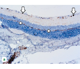
Актуальність. Діабетична ретинопатія (ДР) є основною причиною втрати зору серед пацієнтів з цукровим діабетом (ЦД). Одним із ключових механізмів її прогресування є нейродегенерація, що супроводжується зниженням рівня нейрофіламентів (NF) у сітківці. Клітинні протеїнкінази беруть участь у регуляції апоптозу та нейродегенеративних процесів при ДР. Вплив їхньої фармакологічної блокади на рівень NF у сітківці потребує подальшого вивчення. Мета: визначити експресію нейрофіламентів у сітківці при експериментальній діабетичній ретинопатії та вплив на неї фармакологічної блокади клітинних протеїнкіназ сорафенібом. Матеріали та методи. Дослідження проведено на 55 тримісячних самцях щурів лінії Wistar. Експериментальну ДР моделювали шляхом введення стрептозотоцину (50 мг/кг). Тварин розподілили на три групи: контрольна (без лікування), група з введенням інсуліну та група з комбінованим введенням інсуліну та інгібітора протеїнкіназ сорафенібу (50 мг/кг). Уміст NF-H у тканині сітківки визначали методом імуноблотингу, а експресію — імуногістохімічним аналізом. Статистичний аналіз проводили методом ANOVA, вірогідними вважали відмінності при p < 0,05. Результати. При розвитку експериментальної ДР рівень NF-H у сітківці знижувався у 2,2 раза (p < 0,05) порівняно з інтактними тваринами, що свідчило про розвиток нейродегенерації. Введення інсуліну не впливало на рівень NF-H (зниження у 2,0 раза; p < 0,05), тоді як комбіноване застосування інсуліну та сорафенібу сприяло його частковому збереженню (зниження у 1,6 раза; p < 0,05). Крім того, застосування сорафенібу зменшувало гіперфосфорилювання нейрофіламентів, що могло бути пов’язане з його впливом на активність протеїнкіназ. Висновки. Фармакологічна блокада клітинних протеїнкіназ сорафенібом частково запобігає втраті NF-H у сітківці при експериментальній ДР, що вказує на перспективність цього підходу для захисту нейрональних структур від гіперглікемічного ушкодження.
Background. Diabetic retinopathy (DR) is the main cause of vision loss among patients with diabetes mellitus. One of the key mechanisms of its progression is neurodegeneration, which is accompanied by a decrease in the level of neurofilaments (NF) in the retina. Cellular protein kinases are involved in the regulation of apoptosis and neurodegenerative processes in DR. The effect of their pharmacological blockade on the level of NF in the retina requires further study. The purpose is to determine the expression of neurofilaments in the retina in experimental diabetic retinopathy and the effect on it of pharmacological blockade of cellular protein kinases with sorafenib. Materials and methods. The study was performed on 55 three-month-old male Wistar rats. Experimental DR was modelled by administration of streptozotocin (50 mg/kg). Animals were divided into three groups: controls (no treatment), insulin group and group with combined administration of insulin and protein kinase inhibitor sorafenib (50 mg/kg). The content of NF-H in the retinal tissue was determined by immunoblotting, and its expression — by immunohistochemical analysis. Statistical analysis was performed by ANOVA, differences at p < 0.05 were considered significant. Results. With the development of experimental DR, the level of NF-H in the retina decreased by 2.2 times (p < 0.05) compared to intact animals, indicating the development of neurodegeneration. Insulin administration had no effect on NF-H levels (2.0-fold decrease; p < 0.05), whereas the combined use of insulin and sorafenib contributed to its partial preservation (1.6-fold decrease; p < 0.05). In addition, sorafenib administration reduced neurofilament hyperphosphorylation, which could be related to its effect on protein kinase activity. Conclusions. Pharmacological blockade of cellular protein kinases with sorafenib partially prevents the loss of NF-H in the retina in experimental DR, indicating the prospects of this approach for the protection of neuronal structures from hyperglycemic damage.
діабетична ретинопатія; нейродегенерація; нейрофіламенти; клітинні протеїнкінази; сорафеніб
diabetic retinopathy; neurodegeneration; neurofilaments; cellular proteinkinases; sorafenib
Для ознакомления с полным содержанием статьи необходимо оформить подписку на журнал.
- Ntikoudi M, Farmaki TM, Tziomalos K. Dopamine: A New Player in the Pathogenesis of Diabetic Retinopathy? Int J Mol Sci. 2024 Dec 8;25(23):13196. doi: 10.3390/ijms252313196. PMID: 39684908; PMCID: PMC11642112.
- Bianco L, Arrigo A, Aragona E, Antropoli A, Berni A, Saladi–no A, Battaglia Parodi M, Bandello F. Neuroinflammation and neurodegeneration in diabetic retinopathy. Front Aging Neurosci. 2022 Aug 16;14:937999. doi: 10.3389/fnagi.2022.937999. PMID: 36051309; PMCID: PMC9424735.
- Sohn EH, van Dijk HW, Jiao C, Kok PH, Jeong W, Demir–kaya N, Garmager A, Wit F, Kucukevcilioglu M, van Velthoven ME, DeVries JH, Mullins RF, Kuehn MH, Schlingemann RO, Sonka M, Verbraak FD, Abràmoff MD. Retinal neurodegeneration may precede microvascular changes characteristic of diabetic retinopathy in diabetes mellitus. Proc Natl Acad Sci U S A. 2016 May 10;113(19):E2655-64. doi: 10.1073/pnas.1522014113. Epub 2016 Apr 25. PMID: 27114552; PMCID: PMC4868487.
- Wang W, Lo ACY. Diabetic Retinopathy: Pathophysiology and Treatments. Int J Mol Sci. 2018 Jun 20;19(6):1816. doi: 10.3390/ijms19061816. PMID: 29925789; PMCID: PMC6032159.
- Tien T, Zhang J, Muto T, Kim D, Sarthy VP, Roy S. High Glucose Induces Mitochondrial Dysfunction in Retinal Müller Cells: Implications for Diabetic Retinopathy. Invest Ophthalmol Vis Sci. 2017 Jun 1;58(7):2915-2921. doi: 10.1167/iovs.16-21355. PMID: 28586916; PMCID: PMC5460955.
- Sasaki M, Ozawa Y, Kurihara T, Kubota S, Yuki K, Noda K, Kobayashi S, Ishida S, Tsubota K. Neurodegenerative influence of oxidative stress in the retina of a murine model of diabetes. Diabetologia. 2010 May;53(5):971-9. doi: 10.1007/s00125-009-1655-6. Epub 2010 Feb 17. PMID: 20162412; PMCID: PMC2850533.
- Bek T. Diameter Changes of Retinal Vessels in Diabetic Retinopathy. Curr Diab Rep. 2017 Aug 8;17(10):82. doi: 10.1007/s11892-017-0909-9. PMID: 28791532.
- Callan A, Jha S, Valdez L, Tsin A. Cellular and Molecular Mechanisms of Neuronal Degeneration in Early-Stage Diabetic Retinopathy. Curr Vasc Pharmacol. 2024;22(5):301-315. doi: 10.2174/0115701611272737240426050930. PMID: 38693745.
- Reis A, Mateus C, Melo P, Figueira J, Cunha-Vaz J, Castelo-Branco M. Neuroretinal dysfunction with intact blood-retinal barrier and absent vasculopathy in type 1 diabetes. Diabetes. 2014 Nov;63(11):3926-37. doi: 10.2337/db13-1673. Epub 2014 Jun 19. PMID: 24947354.
- Gafson AR, Barthélemy NR, Bomont P, Carare RO, Durham HD, Julien JP, Kuhle J, Leppert D, Nixon RA, Weller RO, Zetterberg H, Matthews PM. Neurofilaments: neurobiological foundations for biomarker applications. Brain. 2020 Jul 1;143(7):1975-1998. doi: 10.1093/brain/awaa098. PMID: 32408345; PMCID: PMC7363489.
- Devarakonda SS, Basha S, Pithakumar A, L B T, Mukun–da DC, Rodrigues J, K A, Biswas S, Pai AR, Belurkar S, Mahato KK. Molecular mechanisms of neurofilament alterations and its application in assessing neurodegenerative disorders. Ageing Res Rev. 2024 Dec;102:102566. doi: 10.1016/j.arr.2024.102566. Epub 2024 Oct 29. PMID: 39481763.
- Li D, Mielke MM. An Update on Blood-Based Markers of Alzheimer’s Disease Using the SiMoA Platform. Neurol Ther. 2019 Dec;8(Suppl 2):73-82. doi: 10.1007/s40120-019-00164-5. Epub 2019 Dec 12. PMID: 31833025; PMCID: PMC6908531.
- Anwar S, Ahmed A, Sarli V, Hassan I. Editorial: Protein kinase inhibitors in neurodegeneration and cancer targeted therapies. Front Cell Dev Biol. 2024 Apr 18;12:1413293. doi: 10.3389/fcell.2024.1413293. PMID: 38699160; PMCID: PMC11063359.
- Ahmed T, Zulfiqar A, Arguelles S, Rasekhian M, Nabavi SF, Silva AS, Nabavi SM. Map kinase signaling as therapeutic target for neurodegeneration. Pharmacol Res. 2020 Oct;160:105090. doi: 10.1016/j.phrs.2020.105090. Epub 2020 Jul 21. PMID: 32707231.
- Miller WP, Ravi S, Martin TD, Kimball SR, Dennis MD. Activation of the Stress Response Kinase JNK (c-Jun N-terminal Kinase) Attenuates Insulin Action in Retina through a p70S6K1-dependent Mechanism. J Biol Chem. 2017 Feb 3;292(5):1591-1602. doi: 10.1074/jbc.M116.760868. Epub 2016 Dec 13. PMID: 27965359; PMCID: PMC5290937.
- Du Y, Tang J, Li G, Berti-Mattera L, Lee CA, Bartkowski D, Gale D, Monahan J, Niesman MR, Alton G, Kern TS. Effects of p38 MAPK inhibition on early stages of diabetic retinopathy and sensory nerve function. Invest Ophthalmol Vis Sci. 2010 Apr;51(4):2158-64. doi: 10.1167/iovs.09-3674. Epub 2010 Jan 13. Erratum in: Invest Ophthalmol Vis Sci. 2011 Jul;52(8):6057. Li, Guanyuan [corrected to Li, Guangyuan]. PMID: 20071676; PMCID: PMC2868413.
- Li J, Chen K, Li X, Zhang X, Zhang L, Yang Q, Xia Y, Xie C, Wang X, Tong J, Shen Y. Mechanistic insights into the alterations and regulation of the AKT signaling pathway in diabetic retinopathy. Cell Death Discov. 2023 Nov 17;9(1):418. doi: 10.1038/s41420-023-01717-2. PMID: 37978169; PMCID: PMC10656479.
- Guan R, Kang Z, Li L, Yan X, Gao T. PIK3CA regulates development of diabetes retinopathy through the PI3K/Akt/mTOR pathway. PLoS One. 2024 Jan 9;19(1):e0295813. doi: 10.1371/journal.pone.0295813. PMID: 38194422; PMCID: PMC10775978.
- Dabbs D. Diagnostic Immunohistochemistry, 6th Edition Theranostic and Genomic Applications. 2021. 1000 p.
- Schirmer L, Antel JP, Brück W, Stadelmann C. Axonal loss and neurofilament phosphorylation changes accompany lesion development and clinical progression in multiple sclerosis. Brain Pathol. 2011 Jul;21(4):428-40. doi: 10.1111/j.1750-3639.2010.00466.x.
- De Paoli LF, Kirkcaldie MTK, King AE, Collins JM. Neurofilament heavy phosphorylated epitopes as biomarkers in ageing and neurodegenerative disease. J Neurochem. 2025 Feb;169(2):e16261. doi: 10.1111/jnc.16261.
- Völgyi B, Bloomfield SA. Axonal neurofilament-H immunolabeling in the rabbit retina. J Comp Neurol. 2002 Nov 18;453(3):269-79. doi: 10.1002/cne.10392.
- Khalil M, Teunissen CE, Otto M, Piehl F, Sormani MP, Gattringer T, Barro C, Kappos L, Comabella M, Fazekas F, Petzold A, Blennow K, Zetterberg H, Kuhle J. Neurofilaments as biomarkers in neurological disorders. Nat Rev Neurol. 2018 Oct;14(10):577-589. doi: 10.1038/s41582-018-0058-z. PMID: 30171200.
- Barry DM, Stevenson W, Bober BG, Wiese PJ, Dale JM, Barry GS, Byers NS, Strope JD, Chang R, Schulz DJ, Shah S, Calcutt NA, Gebremichael Y, Garcia ML. Expansion of neurofilament medium C terminus increases axonal diameter independent of increases in conduction velocity or myelin thickness. The Journal of Neuroscience. 2012 May 2;32(18):6209-19. doi: 10.1523/jneurosci.0647-12.2012
- Yuan A, Rao MV, Veeranna, Nixon RA. Neurofilaments and neurofilament proteins in health and disease. Cold Spring Harbor Perspectives in Biology. 2017 Apr;9(4). doi: 10.1101/cshperspect.a018309.
- Khalil M, Pirpamer L, Hofer E, Voortman MM, Barro C, Leppert D, Benkert P, Ropele S, Enzinger C, Fazekas F, Schmidt R, Kuhle J. Serum neurofilament light levels in normal aging and their association with Morphologic Brain Changes. Nature Communications. 2020 Feb 10;11(1). doi: 10.1038/s41467-020-14612-6.
- Garzone D, Finger RP, Mauschitz MM, Santos ML, Brete–ler MM, Aziz NA. Neurofilament light chain and retinal layers’ determinants and association: A population‐based study. Annals of Clinical and Translational Neurology. 2022 Mar 4;9(4):564-9. doi: 10.1002/acn3.51522.
- Petzold A, Junemann A, Rejdak K, Zarnowski T, Thaler S, Grieb P, et al. A novel biomarker for retinal degeneration: Vitreous Body Neurofilament proteins. Journal of Neural Transmission. 2009 Sept 22;116(12):1601-6. doi: 10.1007/s00702-009-0316-8.
- Petzold A. The 2022 lady Estelle Wolfson Lectureship on Neurofilaments. Journal of Neurochemistry. 2022 Sept 19;163(3):179-219. doi: 10.1111/jnc.15682.
- Sihag RK, Inagaki M, Yamaguchi T, Shea TB, Pant HC. Role of phosphorylation on the structural dynamics and function of types III and IV intermediate filaments. Experimental Cell Research. 2007 Jun;313(10):2098-109. doi: 10.1016/j.yexcr.2007.04.010.
- Jingi AM, Tankeu AT, Ateba NA, Noubiap JJ. Mechanism of worsening diabetic retinopathy with rapid lowering of blood glucose: The synergistic hypothesis. BMC Endocrine Disorders. 2017 Oct 10;17(1). doi: 10.1186/s12902-017-0213-3.
- Hasskarl J. Sorafenib: Targeting multiple tyrosine kina–ses in cancer. Recent Results in Cancer Research. 2014;145-64. doi: 10.1007/978-3-642-54490-3_8.
- Kwong JM, Caprioli J, Piri N. RNA binding protein with multiple splicing: A new marker for retinal ganglion cells. Investigative Opthalmology & Visual Science. 2010 Feb 1;51(2):1052. doi: 10.1167/iovs.09-4098.