Архив офтальмологии Украины Том 13, №1, 2025
Вернуться к номеру
Стан рогівки як предиктор успішності факоемульсифікації катаракти
Авторы: Скрипник Р.Л., Гребень Н.К., Скрипниченко І.Д.
Національний медичний університет імені О.О. Богомольця, м. Київ, Україна
Рубрики: Офтальмология
Разделы: Клинические исследования
Версия для печати
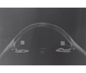
Актуальність. Стан рогівки є важливим фактором, що визначає успішність факоемульсифікації катаракти (ФЕК). Зміни в ендотеліальному шарі, передопераційний рогівковий набряк та інші морфологічні особливості рогівки можуть впливати на швидкість відновлення зору та ризик післяопераційних ускладнень. Мета: оцінити вплив стану рогівки на результати ФЕК шляхом вивчення товщини рогівки та щільності ендотеліальних клітин, визначити її роль у прогнозуванні успішності операції. Матеріали та методи. У дослідженні взяли участь 76 пацієнтів із віковою катарактою, яких було розподілено на дві групи: група 1 — 38 пацієнтів з нормальною рогівкою без ознак ендотеліальної дисфункції, група 2 — 38 пацієнтів з патологічними змінами рогівки, включно зі зниженою щільністю ендотеліальних клітин та передопераційним набряком. Передопераційне обстеження включало візометрію, авторефрактометрію, пахіметрію, ендотеліальну мікроскопію, тонометрію, оптико-когерентну томографію (ОКТ) переднього відділу ока. Післяопераційний моніторинг проводився на 1-й день, а також через 1, 3 і 6 місяців після операції. Результати. Через добу після операції гострота зору була вищою у групі 1 (0,74 ± 0,12) порівняно з групою 2 (0,62 ± 0,15; p < 0,05). Через 6 місяців гострота зору у групі 1 досягла 0,94 ± 0,07, у групі 2 — 0,87 ± 0,09, при цьому у 15,8 % пацієнтів 2-ї групи залишалися скарги на знижену контрастну чутливість. Товщина рогівки у групі 2 була більшою перед операцією (574,6 ± 12,3 мкм) порівняно з групою 1 (531,2 ± 10,5 мкм), а на 1-й день після ФЕК набряк був значно вираженішим у групі 2 (595,1 ± 14,7 мкм) на відміну від групи 1 (548,9 ± 11,2 мкм). Через місяць залишковий набряк спостерігався у 21 % пацієнтів групи 2. Щільність ендотеліальних клітин знизилася у післяопераційному періоді у всіх пацієнтів, але у групі 2 втрати були значно більшими (1987 ± 135 клітин/мм² проти 2492 ± 98 клітин/мм² у групі 1; p < 0,05). Внутрішньоочний тиск (ВОТ) після ФЕК тимчасово підвищувався, особливо у групі 2, де на 1-шу добу його середній приріст становив 4,1 ± 0,9 мм рт.ст. (у групі 1 — 3,2 ± 0,7 мм рт.ст.). Через місяць ВОТ нормалізувався у 85 % пацієнтів, але у 8 % пацієнтів 2-ї групи потребував медикаментозної корекції. За результатами ОКТ переднього відділу ока у пацієнтів 1-ї групи зміни товщини рогівки та її структури визначалися в 21 % випадків (8 очей), у пацієнтів 2-ї групи у 73,6 % випадків (28 очей) визначався набряк рогівки, який у 26,3 % випадків (10 очей) був тривалим та стійким. Визначалися збільшення товщини строми рогівки без порушення її епітелію за рахунок набряку кератоцитів, деформація десцеметової оболонки, ендотеліальне потовщення з протрузією ендотеліоцитів, гіперрефлективні зони. Висновки. Передопераційна оцінка стану рогівки є важливим етапом у плануванні ФЕК. Включення визначення рогівкових параметрів до алгоритму прогнозування післяопераційних результатів ФЕК дозволить покращити результати хірургічного лікування катаракти.
Background. The state of the cornea is an important factor determining the success of phacoemulsification. Changes in the endothelial layer, preoperative corneal edema and other morphological features can affect the rate of vision recovery and the risk of postoperative complications. The purpose of the study was to evaluate the influence of corneal state on the results of phacoemulsification by studying corneal thickness and endothelial cell density, to determine its role in predicting the success of the operation. Materials and methods. The study included 76 patients with age-related cataract who were divided into two groups: group 1 — 38 people with normal cornea without signs of endothelial dysfunction, group 2 — 38 participants with pathological corneal changes, including reduced endothelial cell density and preoperative edema. The preoperative examination included visometry, autorefractometry, pachymetry, endothelial microscopy, tonometry, anterior segment optical coherence tomography. Postoperative monitoring was performed on day 1, as well as 1, 3 and 6 months after surgery. Results. One day after the operation, visual acuity was higher in group 1 (0.74 ± 0.12) compared to group 2 (0.62 ± 0.15; p < 0.05). After 6 months, visual acuity in group 1 reached 0.94 ± 0.07, in group 2 — 0.87 ± 0.09, while 15.8 % of patients in the second group still complained of reduced contrast sensitivity. The corneal thickness before surgery was higher in group 2 (574.6 ± 12.3 μm) than in group 1 (531.2 ± 10.5 μm), and on day 1 after phacoemulsification, edema was significantly more pronounced in group 2 (595.1 ± 14.7 μm) as opposed to group 1 (548.9 ± 11.2 μm). One month later, residual edema was observed in 21 % of patients in the second group. Endothelial cell density decreased in the postoperative period in all patients, but the loss was significantly greater in group 2 (1987 ± 135 cells/mm² vs 2492 ± 98 cells/mm² in group 1; p < 0.05). Intraocular pressure after phacoemulsification temporarily increased, especially in group 2, where on the first day, its average increase was 4.1 ± 0.9 mm Hg (in group 1 — 3.2 ± 0.7 mm Hg). After a month, intraocular pressure normalized in 85 % of patients, but in 8 % of participants in the second group, it required medical correction. According to the anterior segment optical coherence tomography data, changes in the corneal thickness and structure were detected in 21 % of cases (8 eyes) in group 1; in group 2, corneal edema was detected in 73.6 % (28 eyes) of cases, it was prolonged and persistent in 26.3 % (10 eyes). There was an increase in the stromal thickness, deformation of the Descemet’s membrane, endothelial thickening with protrusion of endothelial cells, hyperreflective zones. Conclusions. Preoperative assessment of the cornea is an important step in predicting the success of phacoemulsification. The inclusion of corneal parameters in the algorithm for predicting postoperative phacoemulsification results will improve the outcomes of cataract surgery.
факоемульсифікація; катаракта; рогівка; ендотеліальні клітини; прогнозування; оптична когерентна томографія
phacoemulsification; cataract; cornea; endothelial cells; prognosis; optical coherence tomography
Для ознакомления с полным содержанием статьи необходимо оформить подписку на журнал.
- Abdelazeem К, Shafar M, Saleh MGA, Fathalla AM, Soliman W. Relevance of swept-source anterior segment optical cohe–rence tomography for corneal imaging in patients with flap-related complications after Lasik. Cornea. 2019;38:93-97. https//doi.org/10.1097/ICO.0000000000001773.
- Akpek EK, Sheppard JD, Hamm A, Angstmann-Mehr S, Krösser S. Efficacy of a new water-free topical cyclosporine 0.1% solution for optimizing the ocular surface in patients with dry eye and cataract. J Cataract Refract Surg. 2024 Jun 1;50(6):644-650. doi: 10.1097/j.jcrs.0000000000001423. PMID: 38334413; PMCID: PMC11146174.
- Aoki S, Asaoka R, Fujino Y, Nakakura S, Murata H, Kiuchi Y. Comparing corneal biomechanic changes among solo cataract surgery, microhook ab interno trabeculotomy and iStent implantation. Sci Rep. 2023 Nov 6;13(1):19148. doi: 10.1038/s41598-023-46709-5. PMID: 37932377; PMCID: PMC10628136.
- Boden KT, Schlosser R, Reipen L, Seitz B, Januschowski K, Szurman P, et al. The impact of limbus detection, arcus lipoides and limbal vessels on the primary patency of clear cornea incisions in femtosecond laser-assisted cataract surgery. Acta Ophthalmol. 2021 Sep;99(6):e943-e948. doi: 10.1111/aos.14705. Epub 2021 Jan 27. PMID: 33502099.
- Chou WY, Kuo YS, Lin PY. Cataract surgery in patients with Fuchs’ dystrophy and corneal decompensation indicated for Descemet’s membrane endothelial keratoplasty. Sci Rep. 2022 May 19;12(1):8500. doi: 10.1038/s41598-022-12434-8. PMID: 35589882; PMCID: PMC9120518.
- Chong YJ, Azzopardi M, Hussain G, Recchioni A, Gandhe–war J, Loizou C, et al. Clinical Applications of Anterior Segment Optical Coherence Tomography: An Updated Review. Diagnostics. 2024;14(2):122. doi: 10.3390/diagnostics14020122.
- Díez-Ajenjo MA, Luque-Cobija MJ, Peris-Martínez C, Ortí-Navarro S, García-Domene MC. Refractive changes and visual quality in patients with corneal edema after cataract surgery. BMC Ophthalmol. 2022 Jun 2;22(1):242. doi: 10.1186/s12886-022-02452-5. PMID: 35655163; PMCID: PMC9164413.
- Fram NR, Hovanesian JA, Narang P, Narang R, Moloney G, Lin DTC, et al. Radial keratotomy and cataract surgery: A quest for emmetropia. J Cataract Refract Surg. 2023 Aug 1;49(8):898-899. doi: 10.1097/j.jcrs.0000000000001240. PMID: 37482668.
- Han X, Fan Q, Hua Z, Qiu X, Qian D, Yang J. Analysis of corneal astigmatism and aberration in chinese congenital cataract and developmental cataract patients before cataract surgery. BMC Ophthalmol. 2021 Jan 13;21(1):34. doi: 10.1186/s12886-020-01794-2. PMID: 33435913; PMCID: PMC7805192.
- Hansen MM, Bach-Holm D, Kessel L. Biometry and corneal aberrations after cataract surgery in childhood. Clin Exp Ophthalmol. 2022 Aug;50(6):590-597. doi: 10.1111/ceo.14092. Epub 2022 May 16. PMID: 35524701; PMCID: PMC9546075.
- Humayun S, Noor M, Naqvi SAH, Tarar MS, Ishaq M, Yasmeen H. Correlation Between Corneal Keratometry, Central Corneal Thickness and Anterior Chamber Depth as Measured by Wavelight OB 820 Biometer and Wavelight Oculyzer II in Preoperative Cataract Surgery Patients. J Coll Physicians Surg Pak. 2023 Aug;33(8):890-894. doi: 10.29271/jcpsp.2023.08.890. PMID: 37553928.
- Jiang Y, Qin Y, Bu S, Zhang H, Zhang X, Tian F. Distribution and internal correlations of corneal astigmatism in cataract patients. Sci Rep. 2021 Jun 1;11(1):11514. doi: 10.1038/s41598-021-91028-2. PMID: 34075156; PMCID: PMC8169901.
- Knutsson KA, Savini G, Hoffer KJ, Lupardi E, Bertuzzi F, Taroni L, et al. IOL Power Calculation in Eyes Undergoing Combined Descemet Membrane Endothelial Keratoplasty and Cataract Surgery. J Refract Surg. 2022 Jul;38(7):435-442. doi: 10.3928/1081597X-20220601-02. Epub 2022 Jul 1. PMID: 35858193.
- Miao AO, Lin P, Qian D, Xu J, Lu YI, Zheng T. Association Between Endothelial Cell Density and Corneal Thickness in Medium, Short, and Long Eyes of Han Chinese Cataract Patients. Am J Ophthalmol. 2024 Jun;262:10-18. doi: 10.1016/j.ajo.2024.01.036. Epub 2024 Feb 4. PMID: 38316200.
- Moshirfar M, Huynh R, Ellis JH. Cataract surgery and intraocular lens placement in patients with Fuchs corneal dystrophy: a review of the current literature. Curr Opin Ophthalmol. 2022 Jan 1;33(1):21-27. doi: 10.1097/ICU.0000000000000816. PMID: 34743088.
- Nicholson M, Singh VM, Murthy S, Gatinel D, Pereira S, Pradhan A, et al. Current concepts in the management of cataract with keratoconus. Indian J Ophthalmol. 2024 Apr 1;72(4):508-519. doi: 10.4103/IJO.IJO_1241_23. Epub 2024 Feb 23. PMID: 38389251; PMCID: PMC11149527.
- Romano V, Passaro ML, Bachmann B, Baydoun L, Ni Dhubhghaill S, Dickman M, et al. Combined or sequential DMEK in cases of cataract and Fuchs endothelial corneal dystrophy-A systematic review and meta-analysis. Acta Ophthalmol. 2024 Feb;102(1):e22-e30. doi: 10.1111/aos.15691. Epub 2023 May 8. PMID: 37155336.
- Sandali O, El Sanharawi M, Tahiri Joutei Hassani R, Roux H, Bouheraoua N, Borderie V. Early corneal pachymetry maps after cataract surgery and influence of 3D digital visualization system in minimizing corneal oedema. Acta Ophthalmol. 2022 Aug;100(5):e1088-e1094. doi: 10.1111/aos.15060. Epub 2021 Nov 8. PMID: 34750943.
- Singh C, Joshi VP. Cataract surgery in Keratoconus revisi–ted — An update on preoperative and intraoperative considerations and postoperative outcomes. Semin Ophthalmol. 2023 Jan;38(1):57-64. doi: 10.1080/08820538.2022.2112702. Epub 2022 Aug 22. PMID: 35996343.
- Viberg A, Byström B. Incidence of corneal transplantation after challenging cataract surgery in patients with and without corneal guttata. J Cataract Refract Surg. 2021 Mar 1;47(3):358-365. doi: 10.1097/j.jcrs.0000000000000451. PMID: 33086292.
- Yoon YC, Ha M, Whang WJ. Comparison of surgically induced astigmatism between anterior and total cornea in 2.2 mm steep meridian incision cataract surgery. BMC Ophthalmol. 2021 Oct 19;21(1):373. doi: 10.1186/s12886-021-02131-x. PMID: 34666720; PMCID: PMC8524831.
- Zhou S, Chen X, Ortega-Usobiaga J, Zheng H, Luo W, Tu B, Wang Y. Characteristics and influencing factors of corneal higher-order aberrations in patients with cataract. BMC Ophthalmol. 2023 Jul 12;23(1):313. doi: 10.1186/s12886-023-03067-0. PMID: 37438729; PMCID: PMC10337181.

