
Архив офтальмологии Украины Том 13, №1, 2025
Вернуться к номеру
Аналіз результатів вітреоретинальної хірургії ідіопатичних епіретинальних мембран із застосуванням газової тампонади
Авторы: Путієнко О.О., Парій І.О.
Національний університет охорони здоров’я України імені П.Л. Шупика, м. Київ, Україна
Рубрики: Офтальмология
Разделы: Клинические исследования
Версия для печати
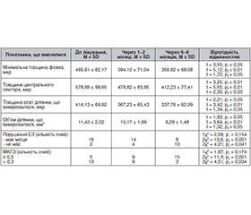
Актуальність. Виникнення ідіопатичних епіретинальних мембран (ЕРМ) — захворювання, яке характеризується клітинною проліферацією із метаплазією клітин, що призводить до формування патологічної фіброзно-клітинної тканини, яка безпосередньо перекриває внутрішню поверхню сітківки. Таке захворювання діагностується у 7–17,8 % осіб, а в осіб старше 80 років — до 28,9 %. На сьогодні найбільш актуальним залишається питання відновлення анатомічного рельєфу сітківки до норми, особливо в далеко розвинутих стадіях процесу, коли спостерігається суттєвий зсув шарів сітківки. Тому виникає питання додаткового впливу на структури сітківки після видалення ЕРМ для її скорішого повного анатомічного відновлення. Мета: оцінити ефективність вітреоретинальної хірургії ідіопатичних ЕРМ із застосуванням газової тампонади. Матеріали та методи. Обстежено 74 пацієнти (85 очей) з ідіопатичними ЕРМ. Згідно з класифікацією за Говетто хворі були розподілені на 4 групи: I група — 12 очей з першою стадією; II група — 30 очей з другою стадією; III група — 25 очей з третьою стадією; IV група — 18 очей з четвертою стадією. Операції виконували калібром 25 або 23G, після виконання субтотальної вітректомії із видаленням задньої гіалоїдної мембрани видаляли епіретинальну мембрану без видалення внутрішньої пограничної мембрани. У I групі операцію завершували на розчині BSS, у II групі — із тампонадою стерильним повітрям, у III та IV групах відповідно — із тампонадою 15% та 20% газоповітряною сумішшю перфторпропану. Результати. У пацієнтів з I стадією через 6–8 місяців спостереження товщина сітківки в центральному секторі (ТСЦС) значуще зменшилась на 53,9 ± 2,7 мкр та в усіх випадках метаморфопсії були відсутні. У пацієнтів із II стадією ТСЦС значуще зменшилась на 77,6 ± 3,4 мкр з вірогідним зменшенням очей з порушенням еліпсоїдної зони (ЕЗ) з 23,3 % до 3,3 %, метаморфопсії залишились тільки на 3 очах (10 %). У пацієнтів з III стадією зменшення ТСЦС в середньому було на 105,2 ± 6,1 мкр з вірогідним зменшенням кількості очей з порушенням ЕЗ з 44 до 16 % та очей з метаморфопсіями з 22 (88,0 %) до 12 (48 %). При IV стадії зменшення ТСЦС було на 166,5 ± 8,2 мкр, порушення ЕЗ з 16 випадків (88,9 %) значуще зменшилось до 8 (44,4 %). Метаморфопсії залишались у більшості випадків — 13 очей (66,7 %). Висновки. Видалення ідіопатичних ЕРМ призводить до значущого покращення гостроти зору, зменшення товщини сітківки, відновлення еліпсоїдної зони та зменшення проявів метаморфопсій протягом 6–8 місяців спостереження на всіх стадіях розвитку цієї патології. Використання стерильного повітря у пацієнтів з ІІ стадією, 15% та 20% газоповітряної суміші перфторпропану як тампонади вітреальної порожнини на очах з ІІІ та IV стадією відповідно дає змогу пришвидшити строк відновлення морфофункціонального стану сітківки та досягти значущого покращення гостроти зору.
Background. Idiopathic epiretinal membranes (ERM) is a disease characterized by cell proliferation and metaplasia, which leads to the formation of pathological fibrous cell tissue that directly overlaps the inner surface of the retina. It occurs in 7–17.8 % of patients, and among people over 80 years of age — in up to 28.9 % of cases. At present, the most urgent issue is to restore the anatomical structure of the retina to normal, especially in advanced stages, when there is a significant shift in the retinal layers. Therefore, there is a question of additional influence on the retinal structures after the removal of ERM for its early full anatomical recovery. Materials and methods. We examined 74 patients (85 eyes) with idiopathic ERM. According to the Govetto classification, patients were divided into 4 groups: group I — 12 eyes with the first stage; group II — 30 eyes with the second stage; group III — 25 eyes with the third stage; group IV — 18 eyes with the fourth stage. The surgeries were performed with 25 or 23G; after subtotal vitrectomy with removal of the posterior hyaloid membrane, the epiretinal membrane was removed without removing the internal limiting membrane. In group I, the operation was finished with BSS, in group II — with sterile air tamponade, in group III and IV, respectively, using tamponade with 15% and 20% perfluoropropane gas-air mixture. Results. In patients with stage I, after 6–8 months of observation, the central retinal thickness (CRT) decreased significantly, by 53.9 ± 2.7 μm, and in all cases, there was no metamorphopsia. In participants with stage II, the CRT decreased significantly, by 77.6 ± 3.4 μm, with a significant reduction in the number of eyes with ellipsoid zone (EZ) disorders from 23.3 to 3.3 %, and metamorphopsia remained in only 3 eyes (10 %). In patients with stage III, the average reduction in the CRT was 105.2 ± 6.1 μm, with a significant decrease in the number of eyes with EZ disorders from 44 to 16 % and eyes with metamorphopsia from 22 (88.0 %) to 12 (48 %). At stage IV, the reduction in the CRT was 166.5 ± 8.2 μm, and the number of EZ disorders significantly decreased from 16 (88.9 %) to 8 cases (44.4 %). Metamorphopsia remained in most cases — 13 eyes (66.7 %). Conclusions. Removal of idiopathic ERM leads to a significant improvement in visual acuity, reduction of retinal thickness, restoration of the ellipsoid zone and a decrease in manifestations of metamorphopsia within 6–8 months of observation at all stages of this pathology. The use of sterile air in patients with stage II, 15% and 20% perfluoropropane gas-air mixture as vitreous cavity tamponade in eyes with stage III and IV, respectively, allows to accelerate the recovery of the morphological and functional state of the retina and to achieve a significant improvement in visual acuity.
ідіопатичні епіретинальні мембрани; вітреоретинальна хірургія; оптична когерентна томографія; газова тампонада; гострота зору
idiopathic epiretinal membranes; vitreoretinal surgery; optical coherence tomography; gas tamponade; visual acuity
Для ознакомления с полным содержанием статьи необходимо оформить подписку на журнал.
- Fraser-Bell S, Guzowski M, Rochtchina E, Wang JJ, Mitchell P. Five-year cumulative incidence and progression of epiretinal membranes: the Blue Mountains Eye Study. Ophthalmology. 2003;110:34-40.
- Ng CH, Cheung N, Wang JJ, Islam AF, Kawasaki R, Meuer SM, et al. Prevalence and risk factors for epiretinal membranes in a multi-–ethnic United States population. Ophthalmology. 2011;118:694-9.
- Kinoshita T, Imaizumi H, Okushiba U, Miyamoto H, Ogino T, Mitamura Y. Time course of changes in metamorphopsia, visual acuity and OCT parameters after successful epiretinal membrane surgery. Investig Ophthalmol Vis Sci. 2016;53:3592-3597.
- Schechet SA, DeVience E, Thompson JT. The effect of internal limiting membrane peeling on idiopathic epiretinal membrane surgery, with a review of the literature. Retina. 2017;37:873-880.
- Azuma K, Ueta T, Eguchi S, Aihara M. Effects of internal limi–ting membrane peeling combined with removal of idiopathic epiretinal membrane: A systematic review of literature and meta-analysis. Retina. 2017;37:1813:1819.
- Chang W, Lin C, Lee C, Sung T, Tung T, Liu J. Vitrectomy with or without internal limiting membrane peeling for idiopathic epiretinal membrane: A meta-analysis. PLoS One. 2017 Jun 16;12(6):e0179105.
- Scheerlinck LME, Van Der Valk R, Van Leeuwen R. Predictive factors for postoperative visual acuity in idiopathic epiretinal membrane: A systematic review. Acta Ophthalmol. 2018;93:203-212.
- Kyuhwan Jang, Daniel Duck-Jin Hwang, Jayoung Ahn, Gisung Son, Ji In Park, Joonhong Sohn. Comparison of the effect of air tamponade versus no tamponade after pars plana vitrectomy for idiopathic epiretinal membrane Scientific Reports. 2021;11:5082. doi: 10.1038/s41598-021-84442-z.
- Govetto A, Lalane RA III, Sarraf D, Figueroa MS, Hubsch–man JP. Insights into epiretinal membranes: presence of ectopic inner foveal layers and a new optical coherence tomography staging scheme. Am J Ophthalmol. 2017;175:99-113.
- Leitritz MA, et al. Early postoperative changes of the foveal surface in epiretinal membranes: Comparison of 23-gauge macular surgery with air vs. balanced salt solution. Graefe’s Arch. Clin. Exp. Ophthalmol. 2014;252:1213-9.
- Lim JW, An TS. Results at 12 months after surgery for epire–tinal membrane: the foveal configurations by optical coherence tomo–graphy. Acta Ophthalmol. 2011;89:e661-e662.
- Hi Y-C, ChuW-L, Chen K-J, Cheng K-C. Morphological Change in Optical Coherence Tomography and Functional Outcomes in Epiretinal Membrane Peeling with or without SF6 Tamponade. Diagnostics. 2024;14:1483. doi: 10.3390/diagnostics14141483.
- Yang HS, Choi S, Kim S, et al. Epiretinal membrane: Correlations among clinical, immunohistochemical, and biochemical features and their prognostic implications. Invest Ophthalmol Vis Sci. 2024;65(14):25. https://doi.org/10.1167/iovs.65.14.25.
- Post M, Cicinelli MV, Zanzottera EC, Marchese A, BandelloF, Coppola M. Prevalence and risk factors of ellipsoid zone damage after pars plana vitrectomy for idiopathic epiretinal membrane. Retina. 2022;42(2):256-264.
- Kim J, Rhee KM, Woo SJ, Yu YS, Chung H, Park KH. Long-term temporal changes of macular thickness and visual outcome after vitrectomy for idiopathic epiretinal membrane. Am J Ophthalmol. 2010;150(5):701-709.e1.