
Архив офтальмологии Украины Том 13, №1, 2025
Вернуться к номеру
Системні фактори ризику прогресування діабетичної ретинопатії при цукровому діабеті 2-го типу
Авторы: Веселовська З.Ф. (1), Панченко Ю.О. (2), Жупан Б.Б. (2), Ковтун М.І. (2), Гуліда А.О. (2)
(1) - Київський медичний університет, м. Київ, Україна
(2) - Національний університет охорони здоров’я України імені П.Л. Шупика, м. Київ, Україна
Рубрики: Офтальмология
Разделы: Клинические исследования
Версия для печати
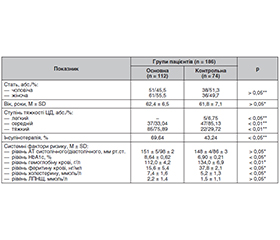
Актуальність. За даними IDF, кількість дорослих із цукровим діабетом (ЦД) у всьому світі у 2021 році досягла 537 млн, що становить 9,8 % населення світу; в Україні кількість пацієнтів з діабетом становила 2,35 млн осіб. Відомо, що діабетична ретинопатія (ДР) є одним із найтяжчих судинних ускладнень діабету та головних причин сліпоти, що вражає близько 40 % хворих на ЦД. Мета: встановити найбільш значущі системні фактори ризику прогресування діабетичної ретинопатії при цукровому діабеті 2-го типу. Матеріали та методи. Проведено аналітичне проспективне когортне дослідження з загальною вибіркою 186 осіб з ЦД2 з української популяції. Обстежені хворі були розподілені на такі групи за станом гіршого ока: основна група — з наявністю будь-якої форми діабетичної ретинопатії (112 осіб), група порівняння — без ознак діабетичної ретинопатії (74 пацієнти). Критеріями включення у дослідження були добровільна інформована згода на участь у дослідженні, вік понад 18 років, наявність верифікованого ЦД2. Вид цукрознижуючої терапії прописував лікар-ендокринолог. Усім пацієнтам було виконано стандартне офтальмологічне обстеження (візометрія, рефрактометрія, дослідження внутрішньоочного тиску та поля зору, гоніоскопія, біомікроскопія, офтальмоскопія, оптико-когерентна томографія, дослідження очного дна на фундус-камері). Серед системних факторів враховувалися показники артеріального тиску — систолічного та діастолічного, показники глікозильованого гемоглобіну, рівень феритину крові та гемоглобіну, рівень холестерину та ЛПНЩ. Статистична обробка результатів дослідження проводилась з використанням ліцензійного пакета програм статистичного аналізу Statistica v6.1. Результати. У більшості хворих на цукровий діабет 2-го типу основної групи (75,89 %) діагностовано тяжку форму захворювання порівняно з 29,72 % групи контролю (р < 0,01). Відповідно до цього на інсулінотерапії перебувало 69,64 % пацієнтів основної групи і лише 43,24 % хворих на ЦД без проявів ретинопатії (р < 0,05). Встановлено, що рівень глікозильованого гемоглобіну у пацієнтів основної групи був значно вищим, ніж у пацієнтів контрольної групи — 8,64 ± 0,62 % проти 6,9 ± 0,21 % (р < 0,05). Рівні гемоглобіну в крові, феритину та холестерину у пацієнтів основної групи були статистично значуще вищими за аналогічні показники контрольної групи (р < 0,05). Рівень ЛПНЩ не мав статистично значущої різниці у пацієнтів обох груп (p > 0,05), проте в основній групі показник був вищим, ніж у контрольній. Висновки. Виникнення діабетичної ретинопатії залежить від тяжкості перебігу цукрового діабету 2-го типу. За нашими даними, у 75,89 % пацієнтів з діабетичною ретинопатією різних стадій діагностовано тяжку форму ЦД порівняно з 29,72 % пацієнтів без проявів ДР (р < 0,01). До системних факторів ризику виникнення ДР можна віднести підвищений рівень глікозильованого гемоглобіну, феритину, гемоглобіну, холестерину крові (р < 0,05).
Background. According to the International Diabetes Federation (IDF), the number of adults with diabetes mellitus worldwide reached 537 million in 2021, which is 9.8 % of the world’s population. In Ukraine, according to the IDF, the number of patients with diabetes was 2.35 million people in 2021. It is known that diabetic retinopathy is one of the most severe vascular complications of diabetes and the main cause of blindness, affecting about 40 % of patients. The purpose was to establish the most significant systemic risk factors for the progression of diabetic retinopathy in type 2 diabetes. Materials and methods. An analytical prospective cohort study was conducted with a total of 186 people with type 2 diabetes from the Ukrainian population. They were divided into the following groups according to the condition of the worse eye: the main group — any form of diabetic retinopathy (112 people) and the comparison group — no signs of diabetic retinopathy (74 patients). The inclusion criteria were voluntary informed consent to participate in the study, age over 18 years, presence of verified type 2 diabetes. The type of glucose-lowering therapy was prescribed by an endocrinologist. All patients underwent a standard ophthalmological examination (visometry, refractometry, intraocular pressure and visual field test, gonioscopy, biomicroscopy, ophthalmoscopy, optical coherence tomography, fundus photography). Systemic factors included were blood pressure — systolic and diastolic, glycosylated hemoglobin, blood ferritin and hemoglobin, cholesterol and low-density lipoprotein levels. Statistical analysis of the study results was performed using the licensed software package Statistica v6.1. Results. Most patients with type 2 diabetes in the main group (75.89 %) were diagnosed with a severe form of disease compared to 29.72 % of controls (p < 0.01). Accordingly, 69.64 % of patients in the main group and only 43.24 % of patients with diabetes without manifestations of retinopathy were on insulin therapy (p < 0.05). It was found that the level of glycosylated hemoglobin in the main group was significantly higher than in controls: 8.64 ± 0.62 % versus 6.90 ± 0.21 % (p < 0.05). The levels of hemoglobin, ferritin and cholesterol in the main group were statistically significantly higher than in the control one (p < 0.05). Low-density lipoprotein level did not have a statistically significant difference in both groups (p > 0.05); however, in the main group, the indicator was higher than in controls. Conclusions. The occurrence of diabetic retinopathy depends on the severity of type 2 diabetes. According to our data, 75.89 % of patients with diabetic retinopathy of various stages were diagnosed with a severe form of diabetes compared to 29.72 % of those without manifestations of diabetic retinopathy (p < 0.01). Systemic risk factors for diabetic retinopathy include elevated levels of glycosylated hemoglobin, ferritin, hemoglobin, and blood cholesterol (p < 0.05).
діабетична ретинопатія; цукровий діабет 2-го типу; фактори ризику
diabetic retinopathy; type 2 diabetes mellitus; risk factors
Для ознакомления с полным содержанием статьи необходимо оформить подписку на журнал.
- Sun H, Saeedi P, Karuranga S, et al. IDF Diabetes Atlas: Global, regional and country-level diabetes prevalence estimates for 2021 and projections for 2045. Diabetes Res Clin Pract. 2022 Jan;183:109119. doi: 10.1016/j.diabres.2021.109119.
- Solomon SD, Goldberg MF. ETDRS Grading of Diabetic Retinopathy: Still the Gold Standard? Ophthalmic Res. 2019;62(4):190-195. https://doi.org/10.1159/000501372.
- Cavan D, Makaroff L, da Rocha Fernandes J, Sylvanowicz M, et al.The Diabetic Retinopathy Barometer Study: Global perspectives on access to and experiences of diabetic retinopathy screening and treatment. Diabetes Res Clin Pract. 2017 Jul;129:16-24. doi: 10.1016/j.diabres.2017.03.023.
- Mohammedi K, Woodward M, Hirakawa Y, et al. Microvascular and Macrovascular Disease and Risk for Major Peripheral Arterial Disease in Patients With Type 2 Diabetes. Diabetes Care. 2016;39(10):1796-1803. doi: 10.2337/dc16-0588.
- Pedro RA, Ramon SA, Marc BB, Juan FB, Isabel MM. Prevalence and relationship between diabetic retinopathy and nephropathy, and its risk factors in the North-East of Spain, a population-based study. Ophthalmic Epidemiol. 2010;17(4):251-65. http://dx.doi.org/10.3109/09286586.2010.498661. PMID: 20642348.
- Karoli R, Fatima J, Shukla V, Garg P, Ali A. Predictors of diabetic retinopathy in patients with type 2 diabetes who have normoalbuminuria. Ann Med Health Sci Res 2013;3(4):536-40. http://dx.doi.org/10.4103/2141-9248.122087. PMID: 24380004.
- International Council of Ophthalmology. ICO Guidelines for Diabetic Eye Care (2017). Availble at: https://icoph.org/eyecare-delivery/diabetic-eye-care/.
- Ting DS, Cheung GC, Wong TY. Diabetic retinopathy: global prevalence, major risk factors, screening practices and public health challenges: a review. Clin Exp Ophthalmol. 2016;44(4):260-77. http://dx.doi.org/10.1111/ceo.12696. PMID: 26716602.
- Solomon SD, Chew E, Duh EJ, et al. Diabetic retinopathy: a position statement by the American Diabetes Association. Diabetes Care. 2017;40:412-8. doi: 10.2337/dc16-2641.
- Foo V, Quah J, Cheung G, et al. HbA1c, systolic blood pressure variability and diabetic retinopathy in Asian type 2 diabetics. J Diabetes. 2017;9(2):200-7. http://dx.doi.org/10.1111/1753-0407.12403. PMID: 27043025.
- Hammes HP, Welp R, Kempe HP, et al. DPV Initiative — German BMBF Competence Network Diabetes Mellitus. Risk Factors for Retinopathy and DME in Type 2 Diabetes-Results from the German/Austrian DPV Database. PLoS One. 2015;10(7):e0132492. http://dx.doi.org/10.1371/journal.pone.0132492. PMID: 26177037.
- Kansal A, Chouhan M, Singh N, Trikha S, Verghese J. Study of anemia in diabetes and its association with diabetic retinopathy. Int J Adv Med. 2017;4:1437-40. http://dx.doi.org/10.18203/2349-3933.ijam20174299.
- American Diabetes Association. Diagnosis and classification of diabetes mellitus. Diabetes Care. 2010;33(1):S62-9. http://dx.doi.org/10.2337/dc10-S062. PMID: 20042775.
- Irace C, Scarinci F, Scorcia V, et al. Association among low whole blood viscosity, haematocrit, haemoglobin and diabetic retinopathy in subjects with type 2 diabetes. Br J Ophthalmol. 2011;95(1):94-8. doi: 10.1136/bjo.2009.172601.