Інформація призначена тільки для фахівців сфери охорони здоров'я, осіб,
які мають вищу або середню спеціальну медичну освіту.

Підтвердіть, що Ви є фахівцем у сфері охорони здоров'я.


Стандартизація надання стоматологічної допомоги як складова успішної клінічної практики

Стандартизація надання стоматологічної допомоги як складова успішної клінічної практики

Oral and General Health Том 5, №2, 2024

Вернуться к номеру

Ураження слизових оболонок ротової порожнини. Клінічні випадки

Авторы: Палатна Л.О., Шпак І.В.
Національний медичний університет імені О.О. Богомольця, м. Київ, Україна

Рубрики: Стоматология

Разделы: Справочник специалиста

Версия для печати


Резюме

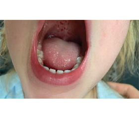
Актуальність. Часто при деяких інфекційних захворюваннях, окрім типових проявів, спостерігаються характерні запальні зміни на слизових оболонках, які можуть імітувати перебіг інших хвороб. Мета дослідження: на прикладі клінічних випадків нагадати стоматологам та лікарям загальної практики про важливість диференціальної діагностики уражень слизової оболонки рота при інфекційних та неінфекційних патологіях. Матеріали та методи. Ми провели емпіричне, описове дослідження 3 клінічних випадків ураження слизової оболонки ротової порожнини у дітей, які проходили стаціонарне лікування в КНП «Київська міська дитяча клінічна інфекційна лікарня») у період 2023–2024 рр. Результати. У першому клінічному випадку описана маніфестація цукрового діабету І типу, що супроводжувалась кандидозом слизової оболонки порожнини рота, який попередньо був розцінений як підозра на кір. Другий клінічний випадок демонструє афтозний стоматит при ВІЛ-інфекції, що попередньо був прийнятий за гострий тонзиліт у дитини. Третій випадок стосувався ентеровірусного везикулярного стоматиту, що за характером елементів нагадував вітряну віспу. Висновки. Ураження слизових оболонок ротової порожнини можуть імітувати прояви поширених інфекційних та неінфекційних хвороб. Тому диференціальна діагностика різних уражень слизової оболонки рота є надзвичайно важливою в різних галузях медицини та стоматології.

Background. Often with some infectious diseases, in addition to the typical manifestations, characteristic inflammatory changes are observed on the mucous membranes, which can imitate the course of other diseases. The purpose was to remind dentists and general practitioners about the importance of differential diagnosis of lesions of the oral mucosa in infectious and non-infectious pathologies on the example of clinical cases. Materials and methods. We conducted an empirical, descriptive study of 3 clinical cases of lesions of the oral mucosa in children who underwent inpatient treatment at the Kyiv City Children’s Clinical Infectious Hospital in 2023–2024. Results. In the first clinical case, the manifestation of type 1 diabetes was described, which was accompanied by oral candidiasis that was previously regarded as a suspicion of measles. The second clinical case demonstrates aphthous ulcers in HIV infection, which was previously mistaken for acute tonsillitis in a child. The third case described enteroviral vesicular stomatitis, which by the nature of the elements resembled chicken pox. Conclusions. Lesions of the oral mucosa can mimic the manifestations of common infectious and non-infectious diseases. Therefore, differential diagnosis of lesions of the oral mucosa is extremely important in various fields of medicine and dentistry.


Ключевые слова

слизова оболонка; ротова порожнина; кандидоз; стоматит; діти

mucous membrane; oral cavity; candidiasis; stomatitis; children


Для ознакомления с полным содержанием статьи необходимо оформить подписку на журнал.


Список литературы

1. Yao H., Zhang Q., Song Q., et al. Characteristics of Oral Mucosal Lesions and Their Association With Socioeconomic Status and Systemic Health: A Cross-Sectional Study of Consecutively Collected Oral Medicine Clinic Data in a Remote Rural Area of China. Front Public Health. 2022;10:897814. doi: 10.3389/fpubh.2022.897814.
2. Dommisch H., Schmidt-Westhausen A.M. The role of viruses in oral mucosal lesions. Periodontol. 2000. Published online February 27, 2024. doi: 10.1111/prd.12553.
3. Panchanadikar N., Balasubramanian S., Nirmal L., et al. Management of oral mucosal lesions in salicylate sensitive stevens-Johnson syndrome — A case report. Indian J Dent Res. 2021;32(4):537-540. doi: 10.4103/ijdr.IJDR_322_20.
4. Mauri-Obradors E., Estrugo-Devesa A., Jané-Salas E., et al. Oral manifestations of Diabetes Mellitus. A systematic review. Med Oral Patol Oral Cir Bucal. 2017;22(5):e586-e594. doi: 10.4317/medoral.21655.
5. Dalipi Z.S., Dragidella F., Dragidella D.K. Oral Manifestations of Exudative Erythema Multiforme in a Patient with COVID-19. Case Rep Dent. 2021:1148945. doi: 10.1155/2021/1148945.
6. Singh A., Jindal A.K., Pilania R.K., et al. Severe Lip Involvement in Kawasaki Disease: A Clinical Oddity. JCR: Journal of Clinical Rheumatology. 2020 Aug;26(5):e149-e150. doi: 10.1097/RHU.0000000000001046.
7. Крамарьов С.О., Надрага О.Б., Буц О.Р. Інфекційні хвороби у дітей. 3-тє вид. К.: ВСВ «Медицина», 2023. С. 279.
8. Husada D., Kusdwijono, Puspitasari D., et al. An evaluation of the clinical features of measles virus infection for diagnosis in children within a limited resources setting. BMC Pediatr. 2020;20(1):5. https://doi.org/10.1186/s12887-020-1908-6. 
9. Mohammadi F., Javaheri M.R., Nekoeian S., et al. Identification of Candida species in the oral cavity of diabetic patients. Curr Med Mycol. 2016;2(2):1-7. https://doi.org/10.18869/acadpub.cmm.2.2.4. 
10. Daniel K., Greenberg R.G., Boutzoukas A., et al. Updated Perspectives on the Diagnosis and Management of Neonatal Invasive Candidiasis. Research and Reports in Neonatology. 2023;13:45-63. https://doi.org/10.2147/RRN.S409779.
11. Lauritano D., Moreo G., Oberti L., et al. Oral Manifestations in HIV-Positive Children: A Systematic Review. Pathogens. 2020;9(2):88. https://doi.org/10.3390/pathogens9020088. 
12. Kerr A.R., Ship J.A. Management strategies for HIV-associated aphthous stomatitis. Am J Clin Dermatol. 2003;4(10):669-680. https://doi.org/10.2165/00128071-200304100-00002. 
13. Charles P.D. Chickenpox vs. Hand, Foot, and Mouth Disease. Emedicinehealth. 2021. https://www.emedicinehealth.com/chickenpox_vs_hand_foot_and_mouth_disease/symptom.htm.
14. Крамарьов С.О., Надрага О.Б., Буц О.Р. Інфекційні хвороби у дітей. 3-тє вид. К.: ВСВ «Медицина», 2023. С. 119.
15. Чернишова Л.I., Волоха А.П., Бондаренко А.В. Інфекційні хвороби у дітей. 3-тє вид. К.: ВСВ «Медицина», 2021. С. 303-304.
16. Bubba L., Broberg E.K., Jasir A., et al. Circulation of non-polio enteroviruses in 24 EU and EEA countries between 2015 and 2017: a retrospective surveillance study. Lancet Infect Dis. 2020;20(3):350-361. doi: 10.1016/S1473-3099(19)30566-3.
17. Шостакович-Корецька Л.Р., Будаєва І.В., Гайдук Т.А. та ін. Ентеровірусна інфекція: Hand-Foot-and-Mouth Disease (HFMD) в Україні. Клінічна інфектологія та паразитологія. 2017. Т. 6(2). С. 185-192. 
18. Malobi I.O. Hand-foot-and-mouth disease. BMJ Best practice. Aug 11, 2023. https://bestpractice.bmj.com/topics/en-gb/685.
19. Miyamoto A., Hirata R., Ishimoto K., et al. An outbreak of hand-foot-and-mouth disease mimicking chicken pox, with a frequent association of onychomadesis in Japan in 2009: a new phenotype caused by coxsackievirus A6. Eur J Dermatol. 2014;24(1):103-104. doi: 10.1684/ejd.2013.2222.

Вернуться к номеру