
Журнал «Травма» Том 26, №2, 2025
Вернуться к номеру
Комбіноване та комплексне лікування пухлин кісток передпліччя
Авторы: Кухарук А.С. (2), Волков В.О. (2), Проценко В.В. (1), Чорний В.С. (2), Солоніцин Є.О. (1)
(1) - ДУ «Інститут травматології та ортопедії НАМН України», м. Київ, Україна
(2) - Національний медичний університет імені О.О. Богомольця, м. Київ, Україна
Рубрики: Травматология и ортопедия
Разделы: Клинические исследования
Версия для печати
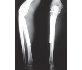
Актуальність. Злоякісні пухлини кісток передпліччя зустрічаються рідко і лишаються проблемою для онкологів-ортопедів. У нашому дослідженні повідомляється про доброякісні місцевоагресивні та злоякісні пухлини кісток передпліччя та різні стратегії хірургічного видалення пухлин і методики реконструкції кісток. Вибір хірургічного втручання при ураженні кісток передпліччя зумовлений не тільки ступенем руйнування кістки пухлиною та нозологічною формою пухлинного процесу, але й здатністю до рецидивування та метастазування пухлини. Проведено аналіз функціональних та онкологічних результатів верхньої кінцівки після комплексного та комбінованого лікування пухлин кісток передпліччя. Мета: показати ефективність комбінованого та комплексного лікування доброякісних місцевоагресивних і злоякісних пухлин кісток передпліччя. Матеріали та методи. Під нашим спостереженням перебували пацієнти (n = 27) із пухлинами кісток передпліччя. Було 17 (62,9 %) пацієнтів із пухлинами променевої кістки та 10 (37,1 %) — із пухлинами ліктьової кістки, 16 (59,2 %) — із гігантоклітинною пухлиною кістки, тоді як 11 (40,8 %) мали злоякісні пухлини. Спостереження у середньому становило 27 (від 6 до 42) місяців. Усім 27 пацієнтам були виконані хірургічні втручання у схемах комбінованого та комплексного лікування. Резекція променевої кістки виконана у 17 хворих, причому найчастіше дистального відділу (13 пацієнтів), проксимального відділу (3), діафіза кістки (1). У 10 хворих виконали резекцію ліктьової кістки, причому найчастіше проксимального відділу ліктьової кістки (5 пацієнтів), дистального відділу (3), діафіза кістки (2). Хірургічні межі пухлини після резекції кістки з пухлиною при морфологічному дослідженні були по краю ураження у 2 пацієнтів. Результати. Післяопераційні ускладнення спостерігалися у 3 (11,1 %) хворих. Функціональні результати верхньої кінцівки після органозберігаючих операцій із приводу пухлин кісток передпліччя за шкалою MSTS становили: після ендопротезування ліктьового суглоба — 74,8 %, після кістково-пластичної операції проксимального відділу ліктьової кістки із застосуванням алотрансплантату та керамічного матеріалу на основі біоактивного скла функція ліктьового суглоба — 96,6 %; після резекції діафіза ліктьової кістки й армованого металоостеосинтезу — 94,5 %; після резекції дистального відділу ліктьової кістки з пухлиною функція променево-зап’ясткового суглоба — 80,4 %; після резекції дистального відділу променевої кістки з пухлиною й автопластики малогомілковою кісткою функція променево-зап’ясткового суглоба — 88,3 %; після резекції дистального відділу променевої кістки з пухлиною й ендопротезування металоцементним імплантом функція променево-зап’ясткового суглоба — 84,2 %; після резекції проксимального відділу променевої кістки з пухлиною функція верхньої кінцівки — 78,5 %; після резекції діафіза променевої кістки й армованого металоостеосинтезу функція кінцівки — 98,4 %. Онкологічні результати: місцевий рецидив пухлини спостерігався у 3 пацієнтів (11,1 %), віддалені метастази — у 3 (7,4 %). Трирічна загальна виживаність хворих становила 82,4 ± 7,6 %, трирічна безрецидивна виживаність — 76,6 ± 8,5 %. Висновки. Рецидив пухлини кістки пов’язуємо з неадекватною відповіддю пухлини на проведені у передопераційний період курси поліхіміотерапії та променевої терапії, а також із нерадикальним видаленням (недодержанням хірургічних меж пухлини під час резекції кістки) пухлини. Віддалені метастази є наслідком рецидиву пухлини після проведеного комплексного та комбінованого лікування та поганим прогностичним фактором виживаності пацієнтів.
Background. Malignant tumors of the forearm bones are rare, and remain a problem for orthopedic oncologists. Our study reports on benign locally aggressive and malignant tumors of the forearm bones, and various strategies for surgical removal of tumors, and bone reconstruction techniques. The choice of surgical intervention in case of the diseased forearm bones is determined not only by the degree of bone destruction by the tumor and the nosological entity of the neoplastic process, but also by the ability of the tumor to recur and metastasize. An analysis of the functional and oncological results of the upper limb after comprehensive and multimodal treatment of tumors of the forearm bones was conducted. Purpose. to show the effectiveness of multimodal and comprehensive treatment of benign locally aggressive and malignant tumors of the forearm bones. Materials and methods. Patients (n = 27) with tumors of the forearm bones were under our observation. There were 17 (62.9 %) patients with radial bone tumors and 10 (37.1 %) patients with ulna tumors, 16 (59.2 %) patients had giant cell tumor of bone, while 11 (40.8 %) had malignant tumors. The average follow-up lasted 27 (range 6 to 42) months. All 27 patients underwent surgical interventions within multimodal and comprehensive treatment regimens. Radial bone resection was performed in 17 patients, at that, most often of distal segment (13 patients), of proximal segment (3 patients), and of diaphysis of bone (1 patient). Ulnar bone resection was performed in 10 patients, at that, most often of proximal ulna (5 patients), of distal ulna (3 patients), and of diaphysis of bone (2 patients). The surgical margin of the tumor, after resection of the bone with the tumor, during morphological examination were along the edge of the lesion in 2 patients. Results. Postoperative complications were observed in 3 (11.1 %) patients. Functionality results of the upper limb after organ sparing surgery for tumors of the forearm bones according to the MSTS scale were: after the elbow joint endoprosthesis replacement the function amounted to 74.8 %, after osteoplastic surgery of the proximal ulna using allograft and ceramic material based on Bioactive Glass, the function of the elbow joint amounted to 96.6 %. After resection of the ulna diaphysis, and effecting reinforced metallic osteosynthesis, it amounted to 94.5 %. After resection of the distal ulna with the tumor, the function of the radiocarpal joint amounted to 80.4 %. After resection of the distal radius with tumor, and effecting fibular bone grafting, the function of the radiocarpal joint amounted to 88.3 %. After resection of the distal radius with tumor, and effecting endoprosthesis replacement using a metal-cement implant, the function of the radiocarpal joint amounted to 84.2 %. After resection of the proximal segment of radius with tumor, the function of the upper limb amounted to 78.5 %. After resection of the radial diaphysis, and effecting reinforced metallic osteosynthesis, the function of the limb amounted to 98.4 %. Oncological results: local tumor recurrence was observed in 3 patients (11.1 %), distant metastases were observed in 2 patients (7.4 %). Three-year overall survival of patients amounted to 82.4 ± 7.6 %, three-year recurrence-free survival amounted to 76.6 ± 8.5 %. Conclusions. Bone tumor recurrence is associated with inadequate tumor response to preoperative courses of polychemotherapy and radiotherapy, as well as with non-radical tumor removal (failure to adhere to surgical tumor margins during bone resection). Distant metastases represent a consequence of tumor recurrence after comprehensive and multimodal treatment, and are an adverse prognostic factor for patient survival.
променева кістка; ліктьова кістка; пухлина; комплексне та комбіноване лікування; функція кінцівки; рецидив; метастаз; виживаність пацієнтів
radius; ulna; tumor; comprehensive and multimodal treatment; limb function; recurrence; metastasis; patient survival