
Международный эндокринологический журнал Том 19, №4, 2023
Вернуться к номеру
Діагностична ефективність інтраопераційного експрес-гістологічного дослідження як інструменту для прийняття рішення про об’єм операції при папілярному раку щитоподібної залози
Авторы: Товкай О.А. (1), Квітка Д.М. (1, 2), Паламарчук В.О. (1), Белемець Н.І. (1), Земсков С.В. (2)
(1) — Український науково-практичний центр ендокринної хірургії, трансплантації ендокринних органів і тканин МОЗ України, м. Київ, Україна
(2) — Національний медичний університет ім. О.О. Богомольця, м. Київ, Україна
Рубрики: Эндокринология
Разделы: Клинические исследования
Версия для печати
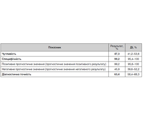
Актуальність. При скринінговому використанні ультразвукової діагностики (УЗД) високої роздільної здатності частка виявлення новоутворень щитоподібної залози (ЩЗ) наближається до 25 % у популяції. Для виявлення карцином ультразвукових критеріїв недостатньо. Для диференціальної діагностики злоякісних новоутворень ЩЗ використовують: УЗД, спіральну комп’ютерну томографію, тонкоголкову аспіраційну пункційну біопсію (ТАПБ), інтраопераційне експрес-гістологічне і патогістологічне дослідження. Деякі автори вважають ТАПБ стандартним методом первинної морфологічної діагностики вузлів ЩЗ. Не завжди є можливість безпечного проведення ТАПБ. Також ТАПБ не є методом стовідсоткової морфологічної ідентифікації первинної пухлини. У таких випадках об’єм операції буде залежати від даних, отриманих при інтраопераційному експрес-гістологічному дослідженні. Аналіз діагностичної ефективності методу експрес-гістології (ЕГ) допоможе у вирішенні питання щодо необхідності його застосування при виборі об’єму оперативного втручання на ЩЗ. Мета роботи: оцінити діагностичну ефективність методу інтраопераційної експрес-гістології у виявлені папілярного раку ЩЗ на солітарних вузлах категорії Bethesda IV, V і метастазів папілярного раку ЩЗ під час операції. Матеріали та методи. Проаналізовано результати інтраопераційних експрес-гістологічних і патогістологічних досліджень, які було виконано в період з 2018 по 2021 роки. У вибірку потрапили матеріали оперативних втручань на ЩЗ при папілярному раку категорій процесу T1ab-2N0-1 і солітарних вузлах категорії Bethesda IV і V за даними ТАПБ. Об’єм вибірки в дослідженні реґіонарного метастазування становив 220 досліджень, по «сірій зоні» — 845 досліджень (категорія Bethesda IV — 465, категорія Bethesda V — 380). Оцінювались випадки розбіжності результатів інтраопераційної ЕГ і кінцевого патогістологічного дослідження. Обробка даних проводилась із застосуванням програми статистичного аналізу EZR v. 3.4.1. Результати. Чутливість методу експрес-гістологічного дослідження в разі виявлення метастазів раку ЩЗ становить 72,2 % при специфічності методу 99,9 %. Діагностична ефективність точність для виявлення метастазів раку ЩЗ становить 89,1 %. При проведенні ЕГ на вузлових утвореннях категорії Bethesda IV чутливість методу становить 8 % при специфічності майже 100 %. Діагностична ефективність ЕГ є вищою при категорії Bethesda V: чутливість — 47 % при маже 100% специфічності. Отримані показники корелюють зі світовими даними. Це свідчить про необхідність подальшого вдосконалення методу інтраопераційної діагностики. Висновки. Діагностична ефективність інтраопераційної ЕГ для виявлення метастазів папілярного раку ЩЗ сягає 89,1 % при 72,2% чутливості та 99,9% специфічності. Діагностична ефективність інтраопераційної ЕГ для верифікації папілярного раку ЩЗ на вузлах категорії Bethesda IV становить 63,9 %, чутливість — 8,2 % і специфічність — 99,6 %. Діагностична ефективність інтраопераційної ЕГ для верифікації папілярного раку ЩЗ на вузлах категорії Bethesda V становить 63,4 %, при цьому чутливість досягає 47,3 %, а специфічність — 99,2 %. Метод інтраопераційної ЕГ дає можливість приймати рішення щодо хірургічної тактики під час оперативного втручання у визначених межах, тому вважаємо за доцільне його подальше використання і вдосконалення.
Background. In case of the screening use of high-resolution ultrasound, the level of detecting thyroid neoplasms is about 25 % in the population. Ultrasound criteria are not enough to detect carcinoma. The following methods are used for the differential diagnosis of malignant thyroid neoplasms: ultrasound scans, computed tomography, fine-needle aspiration (FNA) biopsy, intraoperative express histological and histopathological examination. Some authors consider FNA to be the standard method for primary morphological diagnosis of thyroid nodes. It is not always possible to conduct FNA safely. Also, FNA cannot be a method of 100% morphological identification of a primary tumor. In such cases, the extent of the surgery will depend on the data obtained from the intraoperative express histological examination. Analysis of the diagnostic effectiveness of the express histological examination will help to take a decision on the need for it when choosing the extent of a surgery on the thyroid gland. Aim of the study: to evaluate the diagnostic efficiency of the intraoperative express histological method in the detection of papillary thyroid cancer on solitary nodes (Bethesda IV, V) and metastases of papillary thyroid cancer during surgery. Materials and methods. The results of intraoperative express histological and pathohistological examinations performed at the Ukrainian Scientific and Practical Center for Endocrine Surgery, Transplantation of Endocrine Organs and Tissues from 2018 to 2021 were analyzed. The sample included materials of surgeries on the thyroid gland in case of papillary cancer T1ab-2N0–1 and solitary nodes (Bethesda IV and Bethesda V) according to data obtained using FNA. The sample size in the study of regional metastasis included 220 examinations, 845 examinations in the “gray zone” (Bethesda IV — 465, Bethesda V — 380). Cases of discrepancy between the results of intraoperative express histological and final histopathological examinations were considered. The data was processed using the EZR v. 3.4.1 statistical analysis program. Results. The sensitivity of the express histological examination in case of detecting metastases of thyroid cancer is 72.2 % with the method specificity of 99.9 %. The diagnostic efficiency in detecting metastases of thyroid cancer is 89.1 %. When the express histological examination is used for Bethesda IV nodes, the sensitivity of the method is 8 % with a specificity of almost 100 %. The diagnostic efficiency of the express histological examination is even higher in case of Bethesda V: sensitivity of 47 % with the method specificity of almost 100 %. The values obtained correlate with international data. This testifies to the need for further improvement of the of intraoperative diagnosis method. Conclusions. The diagnostic efficiency of the intraoperative express histological examination for detecting metastases of papillary thyroid cancer in the selected group is 89.1 % with sensitivity of 72.2 % and specificity of 99.9 %. The diagnostic efficiency of intraoperative express histological examination for verification of thyroid cancer in case of Bethesda IV nodes is 63.9 % with sensitivity of 8.2 % and specificity of 99.6 %. The diagnostic efficiency of intraoperative express histological examination for verification of thyroid cancer in Bethesda V nodes is 63.4 % with sensitivity of 47.3 % and specificity of 99.2 %. The intraoperative express histological examination method makes it possible to take decisions on surgical tactics during surgery within the determined limits, so we believe it reasonable to further use and improve it.
щитоподібна залоза, папілярний рак, діагностика, інтраопераційна експрес-гістологія
thyroid gland; papillary cancer; diagnosis; intraoperative express histological examination
Для ознакомления с полным содержанием статьи необходимо оформить подписку на журнал.
- Wong R., Farrell S.G., Grossmann M. Thyroid nodules: diagnosis and management. Med. J. Aust. 2018 Jul 16. 209(2). 92-98. doi: 10.5694/mja17.01204. PMID: 29996756.
- Nabhan F., Dedhia P.H., Ringel M.D. Thyroid cancer, recent advances in diagnosis and therapy. Int. J. Cancer. 2021 Sep 1. 149(5). 984-992. doi: 10.1002/ijc.33690. Epub 2021 May 29. PMID: 34013533.
- Palamarchuk V.O., Kvitka D.M., Mazur O.V. Risk-oriented treatment of papillary thyroid cancer. Clinical Endocrinology and Endocrine Surgery. 2019. (3). 32-38. (in Ukrainian). doi: https://doi.org/10.24026/1818-1384.3(99).2019.170674.
- Hartl D.M., Hadoux J., Guerlain J., Breuskin I., Haroun F., Bidault S., Leboulleux S., Lamartina L. Risk-oriented concept of treatment for intrathyroid papillary thyroid cancer. Best Pract. Res. Clin. Endocrinol. Metab. 2019 Aug. 33(4). 101281. doi: 10.1016/j.beem.2019.05.005. Epub 2019 Jun 4. PMID: 31208873.
- Haddad R.I., Bischoff L., Ball D., Bernet V., Blomain E., Busaidy N.L. et al. Thyroid Carcinoma, Version 2.2022, NCCN Clinical Practice Guidelines in Oncology. J. Natl. Compr. Canc. Netw. 2022 Aug. 20(8). 925-951. doi: 10.6004/jnccn.2022.0040. PMID: 35948029.
- Gal A.A., Cagle P.T. The 100-year anniversary of the description of the frozen section procedure. JAMA. 2005. 294(24). 3135-3136. doi:10.1001/jama.294.24.3135
- Stanciu-Pop C., Pop F.C., Thiry A., Scagnol I., Maweja S., Hamoir E., Beckers A., et al. Controversies regarding the accuracy and limitations of frozen section in thyroid pathology: an evidence-based assessment. Virchows Arch. 2016. 468(2). 135-142. doi: https://doi.org/10.1007/s00428-015-1892-2.
- Sanabria A., Zafereo M., Thompson L.D.R., Hernandez-Prera J.C., Kowalski L.P., Nixon I.J. et al. Frozen section in thyroid gland follicular neoplasms: It's high time to abandon it! Surg. Oncol. 2021 Mar. 36. 76-81. doi: 10.1016/j.suronc.2020.12.005. Epub 2020 Dec 8. PMID: 33316682.
- Estebe S., Montenat C., Tremoureux A., Rousseau C., Bouilloud F., Jegoux F. Limitation of intraoperative frozen section during thyroid surgery. Eur. Arch. Otorhinolaryngol. 2017 Mar. 274(3). 1671-1676. doi: 10.1007/s00405-016-4398-2. Epub 2016 Dec 2. PMID: 27913858.
- Duek S.D., Goldenberg D., Linn S., Krausz M.M., Hershko D.D. The role of fine-needle aspiration and intraoperative frozen section in the surgical management of solitary thyroid nodules. World J. Surg. 2002. 26(12). 1468-1471. doi: https://doi.org/10.1007/s00268-002-6419-3.
- Staubitz J.I., Elmrich I., Musholt P.B., Cámara R.J.A., Watzka F., Dralle H. et al.; Prospective Evaluation Study Thyroid Surgery (PETS) 2 study group. Targeted use of intraoperative frozen-section analysis lowers the frequency of completion thyroidectomy. BJS Open. 2021. 5(2). zraa058. doi: https://doi.org/10.1093/bjsopen/zraa058.
- Basolo F., Ugolini C., Proietti A., Iacconi P., Berti P., Miccoli P. Role of frozen section associated with intraoperative cytology in comparison to FNA and FS alone in the management of thyroid nodules. Eur. J. Surg. Oncol. 2007. 33(6). 769-775. doi: https://doi.org/10.1016/j.ejso.2006.12.004.
- Jonker P., Metman M.J.H., Sondorp L.H.J., Sywak M.S. Intraoperative MET-receptor targeted fluorescent imaging and spectroscopy for lymph node detection in papillary thyroid cancer: novel diagnostic tools for more selective central lymph node compartment dissection. Eur. J. Nucl. Med. Mol. Imaging. 2022 Apr. 49(4). 1080-1081. doi:10.1007/s00259-022-05763-3.
- Tovkai O.A., Kvitka D.M., Palamarchuk V.O., Koza–chuk E.S., Kuts V.V. Assessment of the changes in quality of life of patients with the «low risk» papillary thyroid cancer after surgical treatment. Clinical Endocrinology and Endocrine Surgery. 2022. 4(80). 7-13. DOI: 0.30978/CEES-2022-4-7 (in Ukrainian).