Інформація призначена тільки для фахівців сфери охорони здоров'я, осіб,
які мають вищу або середню спеціальну медичну освіту.

Підтвердіть, що Ви є фахівцем у сфері охорони здоров'я.


Травма та її наслідки
Зала синя Зала жовта

Травма та її наслідки
Зала синя Зала жовта

Журнал «Травма» Том 23, №6, 2022

Вернуться к номеру

Вивчення розподілу напружень моделі заднього міжхребцевого спондилодезу поперекового відділу хребта (імплантат PEEK і дистракційний кейдж)

Авторы: Стогній А.В. (1), П’ятикоп В.О. (1), Яресько О.В. (2), Попсуйшапка К.О. (2), Підгайська О.О. (2), Карпінський М.Ю. (2)
(1) — Харківський національний медичний університет, м. Харків, Україна
(2) — ДУ «Інститут патології хребта та суглобів ім. проф. М.І. Ситенка НАМН України», м. Харків, Україна

Рубрики: Травматология и ортопедия

Разделы: Клинические исследования

Версия для печати


Резюме

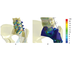
Актуальність. Існує широкий спектр конструкцій і видів матеріалів кейджів для спинномозкових імплантатів, які можуть використовуватися при спондилодезі хребта, але імплантати часто викликають ушкодження замикальної пластини, що може призвести до травми й біомеханічної нестабільності. Існує чотири різних типи матеріалів, які використовують для виготовлення кейджа: металевий, керамічний, полімерний і композитний. Мета: проаналізувати напружено-деформований стан нового міжтілового опорного пристрою і порівняти його з міжтіловою опорою з матеріалу PEEK хребетного блоку L5-S1 і заднього спондилодезу. Матеріали та методи. Була використана інтактна модель таза з крижем і блоком хребців L3-L5. У модель були внесені наступні зміни: міжхребцевий диск L5-S1 був замінений стандартною міжтіловою опорою з матеріалу PEEK. Задній спондилодез L4-S1; міжхребцевий диск L5-S1 був замінений новою міжтіловою опорою. Задній спондилодез L4-S1. Без урахування ваги нижніх кінцівок на верхню поверхню тіла хребця L3 і його суглобові відростки прикладали силу в 422 Н. Результати. Напруження в хребцях L3 і L4 практично не відрізняються від показників моделі в нормі незалежно від типу міжтілової опори. У хребцях L5 і S1 рівень напружень значно перевищує показники моделі в нормі. Імплантат нової конструкції забезпечує значно нижчий рівень напружень на передній поверхні хребця S1 і навколо фіксуючих гвинтів у ньому. Міжтілова опора приймає на себе основне навантаження, про що свідчить величина напружень у ній, яка втричі перевищує максимальний рівень напружень в імплантаті з матеріалу PEEK. Це дозволяє розвантажити елементи транспедикулярної конструкції, що підтверджується низьким рівнем напружень на всіх фіксуючих гвинтах і по всій довжині опорного стрижня. Висновки. Характер розподілу напружень у блоці хребців L3-S1 не змінився порівняно з моделлю PEEK. У тілах хребців L4 і S1 рівень напруження незначно підвищився порівняно з моделлю PEEK, а в хребці L5 знизився. Використання більш жорсткої міжтілової опори дозволило знизити напружений стан на вході фіксуючих гвинтів до кістки хребця S1. Більш високий рівень напруження в самій міжтіловій опорі порівняно з моделлю PEEK не є критичним щодо міцності для металу.

Background. There are a wide variety of designs and materials of spinal implant cages that can be used in spinal fusion, but the implants often cause damage to the locking plate, which can lead to trauma and biomechanical instability. There are four different types of materials used to make a cage: metal, ceramic, polymer, and composite. Goal: to analyze the stress-strain state of the new interbody support device and compare it with the PEEK interbody support of the L5-S1 vertebral block and posterior fusion. Materials and methods. An intact model of the pelvis with the sacrum and the L3-L5 vertebral block was used. The following changes were made to the model: the L5-S1 intervertebral disc was replaced by a standard PEEK interbody support. Posterior fusion of L4-S1; the L5-S1 intervertebral disc was replaced by a new interbody support. Posterior fusion of L4-S1. Without considering the weight of the lower limbs, a force of 422 N was applied to the upper surface of the L3 body and its articular processes. Results. Stresses in the L3 and L4 vertebrae practically do not differ from the values of the normal model, regardless of the type of interbody support. In the L5 and S1 vertebrae, the level of stress significantly exceeds the normal values in the model. The implant of the new design provides a significantly lower level of stress on the front surface of the S1 vertebra and around the fixing screws in it. The interbody support takes on the main load as evidenced by the level of stresses in it, which is three times higher than the maximum stress in the implant made of PEEK material. This allows the elements of the transpedicular structure to be unloaded, which is confirmed by the low level of stress on all the fixing screws and along the entire length of the support rod. Conclusions. The pattern of stress distribution in the L3-S1 vertebral block did not change compared to the PEEK model. In the bodies of the L4 and S1 vertebrae, the stress level slightly increased compared to the PEEK model, and in the L5 vertebra, it decreased. The use of a more rigid interbody support made it possible to reduce the stress state at the entrance of the fixing screws to the bone of the S1 vertebra. The higher level of stress in the interbody support itself, compared to the PEEK model, is not critical in terms of strength for the metal.


Ключевые слова

задній спондилодез; міжтілова опора; напруження

posterior fusion; interbody support; stress


Для ознакомления с полным содержанием статьи необходимо оформить подписку на журнал.


Список литературы

1. Попсуйшапка К.О., Тесленко С.О., Попов А.І., Карпінський М.Ю., Яресько О.В. Дослідження напружено-деформованого стану моделей хребта залежно від обсягу руйнування хребця Th6 і варіанту остеосинтезу. Травма. 2022. Т. 23. № 5. С. 53-64. DOI: https://doi.org/10.22141/1608-1706.5.23.2022.916.
2. Нехлопочин А.С., Нехлопочин С.Н., Карпинский М.Ю., Швец А.И., Карпинская Е.Д., Яресько А.В. Математический анализ и оптимизация конструктивных характеристик стабилизирующих телозамещающих систем для субаксиального цервикоспондилодеза с применением метода конечных элементов. Хирургия позвоночника. 2017. Т. 14. № 1. С. 37-45.
3. Ben-Hatira F., Saidane K., Mrabet A. A finite element modeling of the human lumbar unit including the spinal cord. J. Biomedical Science and Engineering. 2012. 5. 146-152. http://dx.doi.org/10.4236/jbise.2012.53019.
4. Srinivas G.R., Kumar M.N., Deb A. Adjacent Disc Stress Following Floating Lumbar Spine Fusion: A Finite Element Study. Asian Spine J. 2017. 11(4). 538-547. DOI: https://doi.org/10.4184/asj.2017.11.4.538.
5. Zhang Z., Fogel G.R., Liao Z., Sun Y., Sun X., Liu W. Biomechanical evaluation of four surgical scenarios of lumbar fusion with hyperlordotic interbody cage: A finite element study/ Bio-Medical and Materials Engineering. 2018. 29. 485-497. DOI: 10.3233/BME-181004.
6. Tyndyk M.A., Barron V., Mchugh P.E., O’Mahoney D. Generation of a finite element model of the thoracolumbar spine. Acta of bioengineering and biomechanics. Wroclaw University of Technology. 2007. 9(1). 35-46.
7. Aroeira R.M.C., de Melo Pertence A.E., Kemmoku D.T., Greco М. Three-dimensional geometric model of the middle segment of the thoracic spine based on graphical images for finite element analysis. Res. Biomed. Eng. 2017 June. 33(2). 97-104. DOI: http://dx.doi.org/10.1590/2446-4740.08916.
8. Nakashima D., Kanchiku T., Nishida N., Ito S., Ohgi J., Suzuki H. et al. Finite element analysis of compression fractures at the thoracolumbar junction using models constructed from medical images. Experimental and Therapeutic Medicine. 2018. 15. 3225-3230. DOI: 10.3892/etm.2018.5848.
9. Nishida N., Ohgi J., Jiang F., Ito S., Imajo Y., Suzuki H. et al. Finite Element Method Analysis of Compression Fractures on Whole-Spine Models Including the Rib Cage. Hindawi Computational and Mathematical Methods in Medicine. 2019. Vol. 2019. Article ID 8348631. 10 p. https://doi.org/10.1155/2019/8348631.
10. Jalil M.H., Mazlan M.H., Todo М. Biomechanical Comparison of Polymeric Spinal Cages Using Ct Based Finite Element Method. International Journal of Bioscience, Biochemistry and Bioinformatics. 2017. Vol. 7. № 2. doi: 10.17706/ijbbb.2017.7.2.110-117.
11. Sato T., Yonezawa І., Todo М., Takano H., Kaneko K. Biomechanical effects of implant materials on posterior lumbar interbody fusion: comparison of polyetheretherketone and titanium spacers using finite element analysis and considering bone density. J. Biomedical Science and Engineering. 2018. Vol. 11. № 4. Р. 45-59. DOI: 10.4236/jbise.2018.114005.
12. Yahya R., Mazlan M.H., Shuib S., Abdullah A.H. Biomechanical Analysis of Spinal Fusion Cage for Lumbar Vertebrae. International Journal of Recent Technology and Engineering. 2019. Vol. 8. Issue 4. ISSN: 2277-3878. DOI: 10.35940/ijrte.D5206.118419.
13. Lim K.-M., Park T.-H., Lee S.-J., Park S.-J. Design and Biomechanical Verification of Additive Manufactured Composite Spinal Cage Composed of Porous Titanium Cover and PEEK Body. Appl. Sci. 2019. 9. 4258. doi: 10.3390/app9204258.
14. Куценко В.О., Карпінський М.Ю., Суббота І.А., Яресько О.В. Моделювання впливу механічних властивостей міжхребцевих дисків та заднього опорного комплексу на стан поперекових опорно-рухових сегментів. Медицина и... 2007. № 1(16). С. 15-20.
15. Kurowski P.M. Engineering Analysis with SolidWorks Simulation 2012. Published April 11, 2012. 475 р. ISBN: 978-1-58503-710-0.
16. Zienkiewicz O.C., Taylor R.L. The Finite Element Me-thod for Solid and Structural Mechanics. Sixth edition. Butterworth-Heinemann, 2005. 736 p.
17. Kong W.Z., Goel V.K. Ability of the Finite Element Mo-dels to Predict Response of the Human Spine to Sinusoidal Vertical Vibration. Spine. 2003. Vol. 28. № 17. Р. 1961-1967. DOI: 10.1097/01.BRS.0000083236.33361.C5.
18. Vidal-Lesso A., Ledesma-Orozco E., Daza-Benítez L., Lesso-Arroyo R. Mechanical Characterization of Femoral Cartilage Under Unicompartimental Osteoarthritis. Ingenierí Amecánica Tecnología y Desarrollo. 2014. Vol. 4. № 6. 239-246.
19. Boccaccio A., Pappalettere C. Mechanobiology of Fracture Healing: Basic Principles and Applications in Orthodontics and Orthopaedics. Theoretical Biomechanics / Dr Vaclav Klika (Ed.). 2011. Р. 21-48.
20. Mobbs R.J., Loganathan А., Yeung V., Rao P.J. Indications for Anterior Lumbar Interbody Fusion. Orthopaedic Surgery. 2013. 5(3). 153-63. DOI: 10.1111/os.12048.
21. Алямовский А.А. SolidWorks/COSMOSWorks. Инженерный анализ методом конечных элементов. Москва: ДМК Пресс, 2004. 432.

Вернуться к номеру