Резюме
Актуальність. Використання кільцевих фіксаторів спице-стрижневого типу є одним із провідних методів лікування незрощень кісток. Удосконалення конструктивних властивостей системи фіксації є перспективним напрямком поліпшення результатів лікування. Мета дослідження — експериментальне обґрунтування конструкційних удосконалень кільцевих спице-стрижневих фіксаторів. Матеріали та методи. Завданням експерименту було визначити вплив просторової фіксації стрижня в кільцевому спице-стрижневому фіксаторі. Об’єктом дослідження була синтетична кістка Sawbones® із моделлю перелому, який був фіксований кільцевим фіксатором 2 типів. Усі параметри (кількість кілець, спиць, кутів їх перехрестя) були аналогічними. Відмінність двох моделей полягала лише в наявності чи відсутності ребра жорсткості фіксації стрижня. За допомогою універсальної випробувальної машини TIRATEST-2151 визначаються характеристики міцності й деформації матеріалів із максимальним зусиллям до 5 кН. Зразки випробували у трьох режимах: стиск вздовж осі кістки, кручення, згин. У процесі навантаження були записані таблиці, у яких розміщені дані, що реєструвала випробувальна установка (переміщення і сили, що прикладалася). У процесі випробування дослідний зразок, включаючи контрастні точки, фотографували при різних навантаженнях. Зображення обробляли на комп’ютері, використовуючи стандартну систему управління цифровим зображенням. Результати. За отриманими даними, ребро жорсткості при фіксації стрижня кільцевого фіксатора впливало на показники жорсткості системи при усіх видах навантаження. Найбільш чутливим був згин. Так, жорсткість при згині зменшилась на 23 %, при стиску — на 8,5 %. Використання удосконалень фіксації стрижня при лікуванні незрощень кісток гомілки після перелому показало клінічну ефективність. Висновки. Використання ребра жорсткості фіксації стрижня кільцевого спице-стрижневого фіксатора в експерименті довело підвищення показників жорсткості. Найбільш значимим було збільшення параметрів жорсткості при згині. За попередніми даними, застосування удосконалень кільцевих фіксаторів при лікуванні незрощень кісток гомілки після перелому засвідчило клінічну ефективність.
Background. The use of pin and rod ring fixators is one of the leading methods for the treatment of non-union of bones. Improving the structural properties of the fixation system is a promising direction for better treatment results. The purpose of the study was an experimental substantiation of structural improvements of pin and rod ring fixators. Materials and methods. The task of the experiment was to determine the influence of the spatial fixation of the rod in the pin and rod ring fixator. The object of the study was a synthetic Sawbones® bone with a fracture model, which was fixed with a ring fixator of 2 types. All parameters (number of rings, rods, angles of their intersection) were similar. The difference between the two models was only in the presence or absence of a stiffener for fixing the rod. The TIRATEST-2151 universal testing machine determines the strength and deformation characteristics of materials with a maximum force of up to 5 kN. The samples were tested in three modes: compression along the axis of the bone, torsion, bending. During the loading, tables were recorded filled with the data registered by the testing machine (movements and applied forces). During the test, a sample, including contrast points, was photographed under different loads. Images were processed on a computer using a standard digital image management system. Results. According to the obtained data, the stiffener influenced the stiffness indicators of the system under all types of load when fixing the rod of the ring fixator. The most sensitive was the bend. Thus, the stiffness in bending decreased by 23 %, in compression — by 8.5 %. The use of improved rod fixation in the treatment of tibial non-unions after fracture has demonstrated clinical efficacy. Conclusions. The use of a stiffener for fixing the rod of the pin and rod ring fixator in the experiment proved an increase in stiffness indicators. The most significant was an increase in stiffness parameters when bending. According to preliminary data, the use of improved ring fixators in the treatment of non-unions of the tibial bones after a fracture has shown clinical efficacy.
Вступ
Незрощення кісток після переломів, особливо за типом сегментарного дефекту кісткової маси, є актуальною проблемою. Основними методами лікування сегментарних кісткових дефектів є: пластика вільним васкуляризованим трансплантатом малогомілкової кістки, дистракційний остеогенез та використання техніки індукованої мембрани [1–4].
Дистракційна кісткова пластика в лікуванні дефектів і незрощень довгих кісток гомілки за допомогою кільцевих фіксаторів (КФ), що набула поширення по всьому світу, має такі переваги: реконструкція відбувається живою кісткою без шкоди для здорових тканин інших ділянок, із достатньою міцністю й довговічністю, стійкістю до інфекції та довічно відновленою функцією [5–8].
Техніка з використанням КФ малотравматична; створює жорстку фіксацію, достатню для зрощення. Метод має низький ризик глибокого інфікування, економічно вигідний і технічно виконуваний в умовах фінансових та технічних обмежень.
У випадках дистракційного заміщення з використанням дистракційного остеогенезу за Ілізаровим реалізується можливість утворення високоякісної, біологічно нормальної нової кісткової тканини досить великих розмірів, не допускаються небезпечні деформації регенерату у процесі дозрівання. Здійснити дистракційний остеосинтез можна за умов не лише сучасних спеціальних центрів, а й у травматологічних відділеннях районних лікарень. У багатьох випадках незрощень метод більш надійний порівняно з іншими видами пластики.
Усі ці характеристики роблять використання КФ конкурентоспроможним засобом у сучасних умовах. Однак є недоліки здійснення фіксації КФ.
При тривалому застосуванні КФ може розвинутися нестабільність системи «апарат — кістка», зумовлена змінами в системі зовнішньої фіксації або явищами остеопорозу.
Удосконалення технології лікування КФ дозволить поліпшити результати лікування цієї тяжкої патології [9, 10].
Мета роботи — експериментальне обґрунтування конструкційних удосконалень кільцевих спице-стрижневих фіксаторів.
Завдання. В експерименті визначити оптимальну конструкцію просторової фіксації стрижня кільцевих фіксаторів спице-стрижневого типу. Удосконалити технологію лікування незрощень із використанням кільцевих фіксаторів з урахуванням отриманих даних експерименту.
Матеріали та методи
Використання спице-стрижневих кільцевих фіксаторів дозволяє поліпшити властивості класичних спицевих апаратів Ілізарова: використання стрижня замість перехресних спиць знижує ризик можливого розвитку, зменшує вагу та робить його застосування комфортнішим. Недоліками стрижневого компонента кільцевого фіксатора є менша стабільність фіксації та відносна складність вузла з’єднання «стрижень — кільцевий модуль».
Об’єктом дослідження була синтетична кістка Sawbones® із моделлю перелому, який був фіксований кільцевим фіксатором спице-стрижневого типу 2 моделями. Усі параметри (кількість колій, спиць, кутів та площин перелому) були аналогічними. Відмінність двох моделей КФ полягала лише в наявності чи відсутності ребра жорсткості фіксації стрижня (рис. 1, 2).
Дослідження проводились у Національному технічному університеті України «Київський політехнічний інститут імені Ігоря Сікорського», на кафедрі динаміки та міцності машин та опору матеріалів. Використовувалася випробувальна машина TIRATEST-2151 № 48/8.9 (рис. 2).
Зразки випробували у трьох режимах: стиск вздовж осі кістки; кручення; згин.
У процесі випробування дослідний зразок, включаючи контрастні точки, фотографували при різних величинах навантаження. Зображення в цифровому вигляді обробляли на комп’ютері, використовуючи стандартну систему управління цифровим зображенням (рис. 3).
Масштаб визначали шляхом зйомки еталонної мірної плитки в аналогічних умовах. За результатами вимірювань переміщень окремих точок препарату розраховували величини їх взаємних переміщень.
Застосовували спосіб реєстрації переміщень точок біологічних препаратів за допомогою цифрової фотозйомки, що забезпечувало одночасне вимірювання зміщень різних точок біомеханічної системи «відламки кістки — фіксатор». При фотографуванні об’єкта було використано контрастні щодо решти зображення точки (мітки). Контрастні мітки розміщували в площинах кістки. Перед початком випробування проводили фотографування ненавантаженого зразка, що надалі використовували як базове зображення.
Зображення у цифровому вигляді обробляли на комп’ютері, використовуючи стандартну систему керування цифровим зображенням.
У процесі навантаження були записані таблиці, у яких розміщені дані, що реєструвала випробувальна установка. Таблиці містять величини переміщення (мм) і сили (Н), що прикладалася. Щоб виключити вплив на розрахунок жорсткості недійсних результатів (система реєструвала результати і під час того, коли навантаження було знято), необхідно було побудувати графіки залежності сили (Н) від переміщення зразка (мм). Далі виділяли лінійний відрізок графіку залежності «сила — переміщення», за яким розраховувалася жорсткість системи (Н/мм).
Результати та обговорення
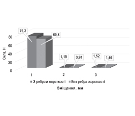
Експериментальні дані порівняння жорсткості фіксації при різних варіантах з’єднання стрижня (із ребром жорсткості і без нього) при дії різних видів навантаження (здавлення по осі; згинання та скручування) подані в табл. 1 і на рис. 5.
Дуже показовою була різниця показників жорсткості Δ при різній фіксації стрижня (рис. 6).
Отримані експериментальні дані свідчать про поліпшення параметрів жорсткості спице-стрижневої системи за допомогою фіксації стрижня з ребром жорсткості. Найбільший ефект був отриманий під час випробування на згин — показник збільшився з 0,91 до 1,19 Н/мм (на 23,53 %); р < 0,05. Це стало підставою для застосування конструктивних поліпшень КФ у хворих із незрощеннями кісток гомілки після переломів. Попередні отримані дані свідчать про наступне.
Застосування в періопераційному знеболюванні нестероїдного протизапального препарату дексалгін із вираженою знеболювальною дією дозволило мінімізувати використання опоїдних препаратів у післяопераційному періоді (лише у 3 хворих основної групи — 16,7 %), що теж вплинуло на функціональні результати.
Отримані наступні результати лікування постраждалих із незрощенням кісток гомілки при застосуванні КФ із запропонованими режимами використання в межах значень оціночної анатомо-функціональної шкали Modified Functional Evaluation System by Karlstrom-Olerud. Описані функції піддавали статистичній обробці та оцінювали за балами. Результати лікування постраждалих із незрощенням кісток гомілки при використанні КФ у запропонованих нами режимах порівнювалися з показниками інших авторів з урахуванням показників оціночної функціональної шкали Modified Functional Evaluation System by Karlstrom-Olerud.
Так, за даними різних авторів [11–13], отримані функціональні результати при лікуванні незрощення великогомілкової кістки коливалися в таких межах: відмінні та хороші — від 37,6 до 84,7 %; погані, незадовільні — від 4,3 та 6,7 до 16,67 %. Отримані нами попередні результи (хороші та відмінні — 77,8 %, незадовільні — 2,8 %) можна порівняти з даними більшості дослідників. Недостатня кількість клінічних спостережень потребує подальшого дослідження.
Висновки
1. Параметри жорсткості спице-стрижневої системи при використанні фіксації стрижня з ребром жорсткості на 23,5 % перевищують такі в системах без ребра жорсткості.
2. Клінічне використання удосконалених спице-стрижневих конструкцій КФ дозволило отримати добрі результати.
3. Отримані результати вдосконаленого остеосинтезу КФ слід вважати обнадійливими, проте ця проблема потребує подальшого вивчення.
Конфлікт інтересів. Автори заявляють про відсутність конфлікту інтересів та власної фінансової зацікавленості при підготовці даної статті.
Інформація про фінансування. Робота виконана у рамках НДР «Теорія та методика ефективного лікування постраждалих з порушенням регенерації тканин». Державний реєстраційний номер 0121U108114. Дата реєстрації: 10.02.2021.
Отримано/Received 03.08.2022
Рецензовано/Revised 15.08.2022
Прийнято до друку/Accepted 23.08.2022
Список литературы
1. Borzunov D.Y., Kolchin S.N., Malkova T.A. Role of the Ilizarov non-free bone plasty in the management of long bone defects and nonunion: problems solved and unsolved. World Journal of Orthopaedics. 2020. 11. 6. 304-318. https://doi.org/10.5312/wjo.v11.i6.304
2. Birch J.G. A brief history of limb lengthening. Journal of Pediatric Orthopaedics. 2017. 37. 2. S1-S8. https://doi.org/10.1097/BPO.0000000000001021
3. Van Niekerk A.H., Birkholtz F.F., de Lange P., Tetsworth K., Hohmann E. Circular external fixation and cemented PMMA spacers for the treatment of complex tibial fractures and infected nonunions with segmental bone loss. Journal of Orthopaedic Surgery. 2017. 25. 2. 230949901771624. https://doi.org/10.1177/2309499017716242
4. Guerado E., Caso E. Challenges of bone tissue engineering in orthopaedic patients. World Journal of Orthopedics. 2017. 8. 2. 87-98. https://doi.org/10.5312/wjo.v8.i2.87
5. Abulaiti A., Liu Y., Cai F. et al. Bone Defects in Tibia Managed by the Bifocal Versus Trifocal Bone Transport Technique. Research Square. 2021. DOI: 10.21203/rs.3.rs-985713/v1.
6. Jiang Q., Huang K., Liu Y., Chi G. Using the Ilizarov technique to treat limb shortening after replantation of a severed lower limb: a case report. Ann. Transl. Med. 2020 Aug. 8(16). 1025. doi: 10.21037/atm-20-5316.
7. Veselý R., Procházka V. Kalusdistrakce v léčení poúrazových defektů femuru a tibie. Acta Chir. Orthop. Traumatol. Cech. 2016. 83(6). 388-392. Czech. PMID: 28026734.
8. Liu Y., Yushan M., Liu Z., Liu J., Ma C., Yusufu A. Complications of bone transport technique using the Ilizarov method in the lower extremity: a retrospective analysis of 282 consecutive cases over 10 years. BMC Musculoskelet Disord. 2020 Jun 6. 21(1). 354. doi: 10.1186/s12891-020-03335-w.
9. Catagni M.A., Azzam W., Guerreschi F., Lovisetti L., Poli P., Khan M.S., Di Giacomo L.M. Trifocal versus bifocal bone transport in treatment of long segmental tibial bone defects. Bone & Joint Surgery. 2019. 101-B. 2. 162-169. https://doi.org/10.1302/0301-620X.101B2.BJJ-2018-0340.R2
10. Wu Y., Yin Q., Rui Y., Sun Z., Gu S. Ilizarov technique: Bone transport versus bone shortening-lengthening for tibial bone and soft-tissue defects. Journal of Orthopaedic Science. 2018. 23. 2. 341-345. https://doi.org/10.1016/j.jos.2017.12.002
11. Kornah B.A., Safwat H.M., Sultan A.A.M., Abdel-Aal M.A. Journal of Trauma & Treatment. J. Trauma. 2016. 5(4). doi: 10.4172/2167-1222.100033.
12. Ayed A. Al Shahrani, Jaya Shanker Tedla, Irshad Ahmad. Effectiveness of Ilizarov frame fixation on functional outcome in aseptic tibial non-union cases at Abha, Kingdom of Saudi Arabia: an experimental study. J. Taibah Univ. Med. Sci. 2015. 10 (2). 216-221.
13. Aktuglu K., Erol K., Vahabi A. Ilizarov bone transport and treatment of critical-sized tibial bone defects: a narrative review. Journal of Orthopaedics and Traumatology. 2019. 20(1). 1-14. https://doi.org/10.1186/s10195-019-0527-1

/34.jpg)
/35.jpg)
/35_2.jpg)
/36.jpg)
/36_2.jpg)