Інформація призначена тільки для фахівців сфери охорони здоров'я, осіб,
які мають вищу або середню спеціальну медичну освіту.

Підтвердіть, що Ви є фахівцем у сфері охорони здоров'я.


Всесвітній день боротьби із запальними захворюваннями кишечника
день перший
день другий

Коморбідний ендокринологічний пацієнт

Всесвітній день боротьби із запальними захворюваннями кишечника
день перший
день другий

Коморбідний ендокринологічний пацієнт

Международный эндокринологический журнал Том 17, №4, 2021

Вернуться к номеру

Роль тонкоголкової аспіраційної біопсії та гістологічного дослідження в оцінці вузлів щитоподібної залози

Авторы: Xhardo E., Xhemalaj D., Agaçi F. , Kapisyzi P.
University Hospital “Shefqet Ndroqi”, Tirana, Albania

Рубрики: Эндокринология

Разделы: Справочник специалиста

Версия для печати


Резюме

Вузли щитоподібної залози становлять вагому проблему і в основному не мають онкологічної характеристики. Таким чином, вони здебільшого не потребують хірургічного лікування. Ризик малігнізації перебуває в межах від 5 до 15 %. Етапи діагностики злоякісної пухлини повинні включати ретельну клінічну оцінку, лабораторні дослідження, ультразвукове дослідження щитоподібної залози та тонкоголкову аспіраційну пункційну біопсію (ТАПБ). ТАПБ є найважливішим діагностичним інструментом при оцінці вузлів щитоподібної залози. Сьогодні вона розглядається як золотий стандарт діаг­ностики злоякісних новоутворень при раку щитоподібної залози. В огляді автори оцінюють роль ТАПБ та можливості цитологічного дослідження в оцінці вузлів щитоподібної залози. Результати ТАПБ класифікуються як діагностичні (задовільні) або недіагностичні (незадовільні). Незадовільні результати (5–10 %) є наслідком міжклітинних зразків, зазвичай спричинених кістозною рідиною, кров’янистими мазками або неоптимальною підготовкою. Діагностичні мазки зазвичай класифікують на доброякісні, невизначені або злоякісні. Доброякісна цитологія (60–70 %) є негативною для злоякісної пухлини і включає кісти, колоїдний вузол або тиреоїдит Хашимото. Злоякісна цитологія (5 %) майже завжди є позитивною для злоякісної пухлини і містить первинні пухлини щитоподібної залози або нетиреоїдні метастатичні раки. Невизначені або підозрілі зразки (10–20 %) включають атипові зміни, клітини Хартла або фолікулярні ново­утворення. Нова цитологічна класифікація Bethesda включає класифікацію шести категорій факторів ризику. З огляду на зростаючу у всьому світі кількість захворювань на мікроаденоми щитоподібної залози останнім часом рекомендується здійснювати ТАПБ за допомогою УЗД навіть при невеликих (< 1 см) вузлах, якщо вони співвідносяться з підозрілими ультразвуковими ознаками або підозрілими лімфатичними вузлами шиї. ТАПБ — це доступний та надійний діагностичний інструмент, що може бути використаний при відборі пацієнтів на операцію та передопераційну діагностику карцином щитоподібної залози. Автори дійшли висновку, що ТАПБ є золотим стандартом при оцінці вузлів щитоподібної залози і може запобігти багатьом непотрібним операціям. Хибнонегативна цитологія залишається проблемою для клініцистів, які лікують пацієнтів із вузлами щитоподібної залози. Післяопераційна гістологія дає остаточний діагноз. Дослідження підтверджують, що вона має суттєві розбіжності між передопераційною цитологією та післяопераційною гістологією в пацієнтів із вузлами щитоподібної залози. Цитопатологи повинні уточнити свої критерії для виявлення аденоматозної гіперплазії, тиреоїдиту, кістозних уражень або підозрілих вузлів щитоподібної залози, щоб уникнути помилкового діагнозу.

Thyroid nodules present a serious problem, and mostly they do not carry neoplastic characteristics. Thus, they do not need to be surgically treated. The risk of malignancy varies from 5 to 15 %. Steps to diagnose malignancy should include a careful clinical evaluation, laboratory tests, a thyroid ultrasound exam and a fine-needle aspiration biopsy. Fine-needle aspiration biopsy (FNAB) is the most important diagnostic tool in the assessment of thyroid nodules. Today it is considered the gold standard for malignancy diagnosis in thyroid cancer. In this review we evaluate the role of FNAB and post-operative cytology in the evaluation of thyroid nodules. FNA results are classified as diagnostic (satisfactory) or nondiagnostic (unsatisfactory). Unsatisfactory smears (5–10 %) result from hypocellular specimens usually caused by cystic fluid, bloody smears, or suboptimal preparation. Diagnostic smears are conventionally subclassified into benign, indeterminate, or malignant categories. Benign cytology (60–70 %) is negative for malignancy, and includes cysts, colloid nodule, or Hashimoto thyroiditis. Malignant cytology (5 %) is almost always positive for malignancy, and includes primary thyroid tumors or nonthyroid metastatic cancers. Indeterminate or suspicious specimens (10–20 %) include atypical changes, Hurthle cells or follicular neoplasms. The new Bethesda Cytologic Classification has a 6-category classification, subdividing indeterminate further by risk factors. Considering the increasing worldwide incidence of thyroid microadenomas, recently it is recommended to undergo FNAB under ultrasound guidance even in small (< 1cm) nodules if they are correlated with suspicious ultrasonographic features or suspicious neck lymph nodes. FNAB is a cheap and reliable diagnostic tool that can be used in the selection candidates for surgery and pre-operative diagnosis of thyroid carcinomas. It was concluded that FNAB is the gold standard in the evaluation of thyroid nodules and can prevent many unnecessary surgeries. False-negative FNA cytology remains a concern for clinicians treating patients with thyroid nodules. Post-operative histology give the definitive diagnosis and studies confirm that it has a significant discordance between pre-operative cytology and post-operative histology in patients with thyroid nodule. Cytopathologists should strengthen their criteria for the identification of adenomatous hyperplasia, thyroiditis, cystic lesions or suspicious thyroid nodules to avoid misdiagnoses.


Ключевые слова

вузли щитоподібної залози; тонкоголкова аспіраційна пункційна біопсія; цитологічне дослідження; огляд

thyroid nodules; fine-needle aspiration biopsy; post-operative cytology; Bethesda classification; review

Introduction

Thyroid nodules (TNs) are among the common diseases of the endocrine system. Increasing with patient age in both sexes, thyroid nodules are found in up to 20% of adults by palpation and in up to 70 % on sonography and autopsy studies with annual increasing trends worldwide; the malignancy rate is 5–15 % [1, 2]. 
The high prevalence of TNs maybe partly because of the advancements in diagnostic technologies, but it still cannot be explained by traditional risk factors such as sex, age, iodine intake, and radiation exposure. Thyroid nodules are a clinical condition that continue to be a problem and still today presents difficulties and triggers debates related to its diagnosis and treatment. Many diagnostic methods have been proposed to differentiate benign nodules from malignant lesions. High-resolution sonography is commonly used to evaluate the thyroid gland, and some sonographic features may help distinguish benign from malignant nodules.
Sonographic features that increase the likelihood of a malignant nodule include size, marked hypoechogenicity, irregular margins, interval growth and the presence of microcalcifications, lymphadenopathy, and local invasion of adjacent structures [3, 4]. The presence of at least 2 suspicious ultrasound criteria reliably identifies 85 % to 93 % of thyroid malignancies [5].
Within the last 20 years, fine-needle aspiration biopsy (FNAB), new radionuclide agents and sensitive ultrasonography have been widely used in the diagnosis of thyroid nodules [6, 7]. In the evaluation of thyroid nodules detected by physical examination and ultrasonography, FNAB should be preferred priorly because of the low cost and the most accurate results [8].
Nowadays, FNAB is recommended as the first and most important step in the management of nodular thyroid disease. 
The FNAB method was first described by Martin and Ellis in 1930, who used an 18-gaugeneedle aspiration technique. The accuracy of the cytological analysis with this method varies between 50–97 % according to the biopsy and the experience of the cytopathology expert.

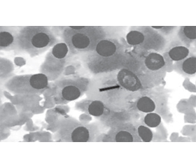
Fine-needle aspiration (FNA) biopsy of thyroid nodules

FNA biopsy was introduced by skandinavian investigators in the early 1960’s and this technique came into widespread use in North America in the 1980’s. Now this technique has become the gold standard diagnostic test (American Thyroid Association (ATA) Grade A recommendation) for initial evaluation of a thyroid nodule, along with a serum TSH level [8].
Most endocrinologists routinely perform FNA biopsy (usually a FNA under ultrasonographic guidance), for the diagnosis of thyroid nodules or for the follow-up surveillance of patients with thyroid cancer.
As the name indicates, the biopsy technique uses aspiration to obtain cells or fluid from a mass. In contrast to percutaneous large needle biopsy, which obtains tissue specimens and requires histologic fixation, aspiration biopsy offers cytologic examination of the specimen. 
Usually, 3 to 6 aspirations and frequently, 8 to 10 slides are made for each nodule [10]. 
The procedure can be palpation-directed or ultrasound-guided and aspirates should be obtained from different portions of the nodule, preferably peripheral areas, in an organized and sequential manner.
Thyroid FNA biopsy, particularly under ultrasound (US-FNA), is very safe. The procedure will take several minutes and no serious complications such as tumor seeding, nerve damage, tissue trauma, or vascular injury have been reported [11, 12].
Patient use of anticoagulants or salicylates does not preclude FNA biopsy. Apart slight pain and some skin discoloration at the aspiration site(s),other complications are extremely rare.
The conventional cytologic diagnosis includes 4 categories: benign (negative), suspicious (indeterminate), malignant (positive), or unsatisfactory (nondiagnostic). 
The new Bethesda Classification has 6 categories, further expanding the indeterminate cytologies.
Aspirates obtained from multinodular goiters, benign microfollicular adenoma, or normal thyroid are referred as colloid nodules and show loosely cohesive sheaths of follicular epithelium, colloid, blood, and rare macrophages (fig. 1).
Benign cytology (60–70 %) is negative for malignancy, and apart multinodular goiters and benign microfollicular adenoma include cysts, colloid nodule, Hashimoto’s thyroiditis and subacute thyroiditis.
Hashimoto’s thyroiditis has a fairly characteristic pattern on FNA smears, showing hypercellularity with lymphocytes, Hürthle cells, and minimal or no colloid.
Subacute (granulomatous) thyroiditis is a rare condition with a benign aspirate. Typically, the smear shows multinucleated giant cells, epithelioid histiocytes, and scattered inflammatory cells.
A benign (negative) cytologic diagnosis is reported for 50 to 90 % of the specimens [13, 14]. 
20 % of FNA cytologic specimens may be suspicious for malignancy (indeterminate) [15, 16]. Decision-making in the “indeterminate” category possesses the greatest challenge for the clinical endocrinologist.
Unsatisfactory smears (5–10 %) result from hypocellular specimens usually caused by cystic fluid, bloody smears, or suboptimal preparation. 
A malignant (positive) cytologic diagnosis varies from 1 to 10 %. Malignant cytology is almost always positive for malignancy, and includes primary thyroid tumors or nonthyroid metastatic cancers.
Papillary thyroid carcinoma (PTC) is the most common malignancy, characterized by increased cellularity, sheets of cells, and typical nuclear abnormalities (fig. 2).
When a positive result for malignancy is obtained, the predictive value of the test is almost 100 %. 
Ultrasound-guided FNA has been found to be superior to palpation-guided FNA due to reduced inadequate sampling and need for repeat biopsy with inadequate sample rates of 14–21 % versus 32–50 %, respectively [17, 18].

False-negative and false-positive rate of thyroid nodule FNA

Evaluation of accuracy of FNA is important due to its clinical implications. 
False-negative results mean missed malignancy. False-negative rates generally vary from 1.5 to 11.5 % [19]. Most of the studies report that false-negative rates of FNA for thyroid nodules are less than 5 % [20–22]. However, higher rates (varying from 7.5 to 21 %) have also been published in other study series [23–25].
The false-negative rate is defined as the percentage of patients with benign cytology in whom malignant lesions are later confirmed on post-operative histology after thyroidectomy. The frequency of false-negative cytologic diagnosis depends on the number of patients who subsequently have surgery and histologic review.
Inadequate or improper sampling accounts for some false-negative errors. Nodules smaller than 1 cm may be too small for accurate needle placement, and nodules larger than 4 cm are too large to allow proper sampling from all areas, thereby increasing the likelihood of misdiagnosis. 
FNA biopsy of thyroid lymphomas may produce lymphocytes that can be interpreted as Hashimoto’s thyroiditis, accounting for a false negative diagnosis. A false positive diagnosis indicates that a patient with a malignant FNA result was found on postsurgical histologic examination to have benign lesions. False-positive rates vary from 0 to 8 % [24, 25].
Hashimoto’s thyroiditis is probably the most common cause of false positive cytology. Misclassification of follicular and Hürthle cell adenomas as papillary carcinomas accounts for other errors. False-negative FNA cytology is especially problematic, as it can result in delayed treatment, which may adversely affect patient outcomes [26].

Causes of false diagnoses

It is not rare that interpretive or sampling errors produce false diagnoses also called misdiagnoses [27–29]. Misdiagnoses can be classified into two categories: “specimen problem”, including sampling error (tumor cells were not aspirated) or a suboptimal specimen (“scant but adequate sampling” or “preparation artifact”), and “interpretation error”, meaning there were overdiagnoses or underdiagnoses by cytologists.
In several studies, the main cause of the false-negative diagnoses was sampling error or specimen problems (86.7 %), while interpretation error led to most of the false-positive diagnoses (80.9 %) [30–32].
Nodule’s size is another factor that can produced false FNA results. In comparison to smaller nodules, the false-negative rate of FNAB seems to be higher for malignancy in larger nodules, especially in nodules measuring larger than 4 cm [32].
To reduce the risk of false-negative findings, multiple passes should be performed in various parts of a large nodule or from different nodules. There is a debate in the literature regarding the role of FNA in the diagnosis of large nodules. Some studies report high false negative results, whereas others note that USG-guided FNA is accurate regardless of nodule size [33].
There was a positive correlation between the nodule size and false negative results. Whereas there is a statistically significant negative correlation between the attendance of cytopathologist and false-negative results [34].
Also, in all the studies regarding this topic, it is evident that an experienced cytopathologist could eliminate most of the false-negative results at the outset.
At the other side, the specimens should be priorly evaluated by cytotechnologist to assess for adequacy of the tissue specimen.
Cytopathologists should re-evaluate their criteria for the identification of adenomatous hyperplasia, thyroiditis and cystic or suspicious lesions to avoid false-positive or false-negative diagnoses.

Diagnostic accuracy

The data of our literature review show that the sensitivity of FNA ranges from 65 to 98 % (mean, 83 %), and specificity ranges from 72 to 100 % (mean, 92 %) [16, 19, 24]. The predictive value of a positive or suspicious cytologic result is approximately 50 % and the overall accuracy rate for cytologic diagnosis varies from 82,5 to 95 % [35, 36].
The studies have shown that the support of ultrasonography (USG) increases the success rate of FNA including specificity and sensitivity [37]. Success of thyroid FNAC depends on skilled aspiration, skilled cytological interpretation and rational analysis of cytological and clinical data. Thyroid FNA in the hands of experienced operators achieves high diagnostic accuracy. To improve accuracy, it is recommended to rebiopsy if cytology is nondiagnostic. In the event that the final result of reaspiration is still insufficient, surgical excision is warranted for most nodules [37].
Evaluation of accuracy of FNA greatly influences the treatment decision. Without other indications, most patients with benign FNAs do not receive surgical resection of thyroid nodules and patients with suspicious or positive for malignancy nodules should go for partial or total thyroidectomy.
Our analysis of the data reveals that FNA provides a good sensitivity, specificity and diagnostic accuracy rate for the evaluation of thyroid nodules, constituting a reliable and effective tool for diagnosis and surgical indication.

FNA guidelines

Guidelines have been published to help improve the adequacy and accuracy of cytology specimens [30]. Current guidelines vary on how to differentiate thyroid nodules into benign or malignant nodules through ultrasound. Recent American Association of Clinical Endocrinologists suggest selection of nodules for FNA on the basis of ultrasound features, whereas the Society of Radiologists in Ultrasound recommends FNA for nodules larger than 1.0 to 1.5 cm in diameter. This issue remains controversial. 
Based on the revised ATA guidelines, FNA is recommended in thyroid nodules larger than 5 mm with suspicious ultrasound features in high-risk patients. This guideline recommend that FNA should be performed regardless of lesion size when patients have a history of neck irradiation, a family history of medullary thyroid cancer or MEN2, extracapsular growth, or metastatic cervical lymph nodes [30]. These last criteria recently are the most used by endocrinologist.

The Bethesda criteria

Terminology of reporting thyroid fine-needle aspiration cytology (FNAC) has varied markedly. Prior to a recently updated cytology classification system for thyroid FNAs, many physicians criticized high false-negative results of thyroid FNAs. Concerns specifically derived from the “indeterminate” category, where rate of malignancy was reported at 40 % [38, 39].
Nowadays, wideworld is used The Bethesda System for Reporting Thyroid Cytopathology (BSRTC) resulted from a conference held at the National Institutes of Health in 2007.
The new Bethesda System for Thyroid Cytopathology offers a 6-category classification that further subdivides the indeterminate category in 3 classes: atypia of undetermined significance (AUS) and follicular lesion of undetermined significance (FLUS); follicular neoplasm; and suspicious for malignancy [40].
This system led to standardization of FNA reports based on six diagnostic categories (DC): 
— DC I = nondiagnostic (ND, Bethesda I).
— DC II = benign (Bethesda II).
— DC III = atypia/follicular lesion of undetermined significance (AUS/FLUS, Bethesda III).
— DC IV = follicular neoplasm/suspicion for a follicular neoplasm (FN/SFN, Bethesda IV).
— DC V = suspicious for malignancy (SM, Bethesda V).
— DC VI = malignant (Bethesta VI) [41].
Each category has an implied cancer risk, which ranges from 0 to 3 % for the “benign” category to virtually 100 % for the “malignant” category. AUS and FLUS have low risk for malignancy, and currently are subjected to molecular testing before surgery. 
This new cytologic classification directly implicit the decision making and clinical management of patients with thyroid nodules. Each cytopathological category is risk stratified for malignancy and corresponds to specific recommendations for patient management [42]. In nondiagnostic or benign categories (DC I and II) it is recommended to repeat FNA under ultrasound guidance and clinical follow-up, respectively. For Atypia of undetermined significance or follicular lesion of undetermined significance the risk of malignancy is 5–15 % and for this reason it is recommended to repeat FNA. Follicular neoplasm or suspicious for a follicular neoplasm cases should go for lobectomy and malignant lesions for near-total or total thyroidectomy.
The routine use of FNA has reduced the number of unnecessary surgical procedures and increased the detection of thyroid cancer at earlier stages [43]. Several studies and literature review analyzing the Bethesda criteria, all concluded that the Bethesda classification system appropriately risk stratified patients preoperatively [43].

Thyroid microcarcinomas

Papillary thyroid microcarcinoma (PTMC) is defined by the World Health Organization (WHO) as malignant lesions < 1 cm in size that are found incidentally. These lesions continue to increase in prevalence worldwide as described by R. Elisei et al. in which microcarcinomas rose from 8% before 1990 to 29 % from 1990 to 2004 [44]. Interestingly, increasing prevalence of thyroid cancer is in large part attributed to increased detection of microcarcinoma [45]. 
As previous studies had noted, PTCs measuring less than 1 cm in diameter are usually incidental and discounted as false-negative by some pathologists [46]. In fact, in several studies, the most common cause of a false-negative diagnosis by FNA is the presence of an unsampled microcarcinoma in the setting of an adenomatous goiter [47]. Finally, thyroid microcarcinomas have been a recent topic of controversy, especially regarding appropriate management. Traditionally, FNA is recommended for nodules greater than 1 cm due to low risk of malignancy in smaller lesions [48]. However, some of these lesions undergo FNA even if they are measuring less than 1 cm, because of suspicious characteristics on ultrasound or suspicious cervical lymph nodes or in patients with a history of radiation to the head and neck region. Ultrasound-guided FNA is effective in the sampling of thyroid cancers that are 1.0 cm or less. Several studies shows that some of these lesions can be clinically significant and earlier detection correlate with a better prognosis.
A retrospective study using data from 1985–1998 in the American College of Surgeons National Cancer Data base [49] demonstrated that papillary thyroid cancers < 1 cm in size were less aggressive than larger tumors with significantly less chance of recurrence and higher survival rates. 
In tumors < 1 cm, there was no difference in recurrence or survival between total thyroidectomy and lobectomy. The opposite result was for tumors > 1 cm where total thyroidectomy, compared to lobectomy, had significantly increased the survival benefit and decreased the risk of recurrence. Thyroid microcarcinoma nowadays are frequently detected at earlier stages but appropriate management of these lesions remains a topic of debate.

Post-operative histology

The final exact diagnosis in the occurrence of thyroid nodules is concluded after partial or total thyroidectomy, through histological examination. Several studies concluded that there is significant discordance between pre-operative cytology and post-operative histology in patients with thyroid nodule [50]. Single cytology sample and even double cytology may miss malignancy 26 and 21 %, respectively [50]. For this reason, a multi-disciplinary approach supported by detailed evidence from history, clinical examination, radiology, and histopathology is required in patients with thyroid nodules.

Discussions

Thyroid diseases are among the most prevalent of medical conditions for this reason it is essential to identify diagnostic tools, especially in the assessment of thyroid nodules. The critical issue in the management of patients with thyroid nodules is to find a way to distinguish preoperatively benign nodules (> 90 %) from cancers [51].
Thyroid FNA biopsy is the most accurate test for determining malignancy, and is an integral part of current thyroid nodule evaluation. FNA has a low false-negative rate for diagnosis of thyroid malignancy; however, controversy exists regarding the accuracy of FNA for nodules smaller than 1 cm or greater than 4 cm. Before 2007 when the The Bethesda System for Reporting Thyroid Cytopathology [40] was published, there was no standard classification and no application of cytological reporting of thyroid nodule FNA results. The Bethesda system classifies the cytological results into 6 categories and presents their respective malignancy rates and approaches to treatment. Since its publication, several centers in different countries have applied the Bethesda classification system to their population, reporting a correlation between their cytological findings and malignancy rates in each category [52]. According to these new criteria, it is recommended that nondiagnostic nodules should be submitted to a second FNA at an appropriate interval. In a series of studies, the malignancy rate obtained in the nondiagnostic category varies from 10–35 %, with 67 % of the cases being microcarcinomas. Clinical follow-up is recommended for nodules classified as benign (Bethesda II) and if significant growth or sonographic changes are observed, another FNA should be performed. According to The Bethesda System, AUS/FLUS nodules (Bethesda III) should be submitted to another FNA after an adequate interval. The majority of them are reclassified into another category after the second FNA and surgical treatment is also recommended. In agreement with the literature AUS/FLUS nodules were reclassified as benign after a second FNA in 42.7–73.1 % of cases, which allows a clinical follow-up approach instead of a surgical procedure.

Conclusions

The accurate diagnosis of thyroid nodules continues to challenge physicians managing patients with thyroid disease.
FNAB is a fast and cost-effective method to evaluate thyroid nodules preoperatively. It is widely recognized as a valuable method in distinguishing neoplastic from non-neoplastic nodules and in identifying patients in need of thyroid surgery.
Nowadays, FNAB is recommended as the first diagnostic test to evaluate thyroid nodules.
This minimally invasive and cost-effective technique is extremely useful in identifying a substantial proportion of thyroid nodules as benign and reducing unnecessary surgery for patients with benign disease.
At the other side, FNAB is a cheap and reliable diagnostic tool used in the selection of candidates for thyroid surgery and preoperative diagnosis of thyroid carcinomas.
The last ATA guidelines recommend that even thyroid microadenomas should be sampled in FNA in the presence of suspicious ultrasound features. 
New Bethesda system has been used to classify thyroid cytology in 6 categories besides presenting malignancy rates and respective approaches.
Several studies evaluating the cytologic-histologic correlation for thyroid diseases concluded that post-operative histology findings can be frequently discordant to pre-operative FNAB but it is important to underline that misdiagnosis can be reduced at maximum and high diagnostic accuracy of FNAB can be archived is in the hands of experienced histopathologist.
 
Received 15.04.2021
Revised 11.05.2021
Accepted 02.06.2021

Список литературы

  1. Kavanagh J., McVeigh N., McCarthy E., Bennett K., Beddy P. Ultrasound-guided fine needle aspiration of thyroid nodules: factors affecting diagnostic outcomes and confounding variables. Acta Radiol. 2017. 58(3). 301-306. doi: 10.1177/0284185116654331. 
  2. Varshney R., Forest V.I., Zawawi F., Rochon L., Hier M.P., Mlynarek A., Tamilia M., Payne R.J. Ultrasound-guided fine-needle aspiration of thyroid nodules: does size matter? Am. J. Otolaryngol. 2014. 35(3). 373-376. doi: 10.1016/j.amjoto.2013.12.005. 
  3. Wong R., Farrell S.G., Grossmann M. Thyroid nodules: diagnosis and management. Med. J. Aust. 2018. 209(2). 92-98. doi: 10.5694/mja17.01204. 
  4. Durante C., Grani G., Lamartina L., Filetti S., Mandel S.J., Cooper D.S. The Diagnosis and Management of Thyroid Nodules: A Review. JAMA. 2018. 319(9). 914-924. doi: 10.1001/jama.2018.0898. 
  5. Singh Ospina N., Iñiguez-Ariza N.M., Castro M.R. Thyroid nodules: diagnostic evaluation based on thyroid cancer risk assessment. BMJ. 2020. 368. 16670. doi: 10.1136/bmj.l6670. 
  6. Cordes M., Schmidkonz C., Horstrup K., Weppler M., Kuwert T. Fine-needle aspiration biopsies of thyroid nodules. Nuklearmedizin. 2018. 57(6). 211-215. doi: 10.3413/Nukmed-0981-18-06. 
  7. Jack G.A., Sternberg S.B., Aronson M.D., Mukamal K.J., Oshin A., Hennessey J.V. Nondiagnostic Fine-Needle Aspiration Biopsy of Thyroid Nodules: Outcomes and Determinants. Thyroid. 2020. 30(7). 992-998. doi: 10.1089/thy.2019.0140.
  8. Haugen B.R., Alexander E.K., Bible K.C., Doherty G.M., Mandel S.J., Nikiforov Y.E., Pacini F. et al. 2015 American Thyroid Association Management Guidelines for Adult Patients with Thyroid Nodules and Differentiated Thyroid Cancer: The American Thyroid Association Guidelines Task Force on Thyroid Nodules and Differentiated Thyroid Cancer. Thyroid. 2016. 26(1). 1-133. doi: 10.1089/thy.2015.0020. 
  9. Choe J., Baek J.H., Park H.S., Choi Y.J., Lee J.H. Core needle biopsy of thyroid nodules: outcomes and safety from a large single-center single-operator study. Acta Radiol. 2018. 59(8). 924-931. doi: 10.1177/0284185117741916.
  10. Mais D.D., Crothers B.A., Davey D.D., Natale K.E., Nayar R., Souers R.J., Blond B.J. et al. Trends in Thyroid Fine-Needle Aspiration Cytology Practices: Results From a College of American Pathologists 2016 Practice Survey. Arch. Pathol. Lab. Med. 2019. 143(11). 1364-1372. doi: 10.5858/arpa.2018-0429-CP. 
  11. Alvarado-Santiago M., Alvarez-Valentin D., Ruiz-Bermudez O., Gonzalez-Sepulveda L., Allende-Vigo M., Santiago-Rodriguez E., Rivas-Tumanyan S. Fine-Needle Thyroid Aspiration Biopsy: Clinical Experience at the Endocrinology Clinics of the University Hospital of Puerto Rico. P. R. Health Sci. J. 2017. 36(1). 5-10. PMID: 28266693; PMCID: PMC5624044.
  12. Crothers B.A., Darragh T.M., Tambouret R.H., Nayar R., Barkan G.A., Zhao C., Booth C.N. et al. Trends in Cervical Cytology Screening and Reporting Practices: Results From the College of American Pathologists 2011 PAP Education Supplemental Questionnaire. Arch. Pathol. Lab. Med. 2016. 140(1). 13-21. doi: 10.5858/arpa.2015-0004-CP.
  13. Padmanabhan V., Barkan G.A., Tabatabai L., Souers R., Nayar R., Crothers B.A. Touch imprint (TI) cytology of needle core biopsies (NCB) in pathology laboratories: A practice survey of participants in the College of American Pathologists (CAP) Non Gynecologic Cytopathology (NGC) Education Program. Diagn. Cytopathol. 2019. 47(3). 149-155. doi: 10.1002/dc.24023. 
  14. Gürkan Dumlu E., Kiyak G., Bozkurt B., Tokaç M., Polat B., Cakir B., Güler G., Kiliç M. Correlation of thyroid fine-needle aspiration with final histopathology: a case series. Minerva Chir. 2013. 68(2). 191-197. PMID: 23612233.
  15. Ha S.M., Kim J.K., Baek J.H. Detection of Malignancy Among Suspicious Thyroid Nodules < 1 cm on Ultrasound with Various Thyroid Image Reporting and Data Systems. Thyroid. 2017. 27(10). 1307-1315. doi: 10.1089/thy.2017.0034. 
  16. Jackson B.S. Controversy regarding when clinically suspicious thyroid nodules should be subjected to surgery: Review of current guidelines. Medicine (Baltimore). 2018. 97(50). e13634. doi: 10.1097/MD.0000000000013634. 
  17. Cesur M., Corapcioglu D., Bulut S., Gursoy A., Yilmaz A.E., Erdogan N., Kamel N. Comparison of palpation-guided fine-needle aspiration biopsy to ultrasound-guided fine-needle aspiration biopsy in the evaluation of thyroid nodules. Thyroid. 2006. 16(6). 555-561. doi: 10.1089/thy.2006.16.555.
  18. Robitschek J., Straub M., Wirtz E., Klem C., Sniezek J. Diagnostic efficacy of surgeon-performed ultrasound-guided fine needle aspiration: a randomized controlled trial. Otolaryngol. Head Neck Surg. 2010. 142(3). 306-309. doi: 10.1016/j.otohns.2009.11.011. 
  19. Giuffrida D., Gharib H. Controversies in the management of cold, hot, and occult thyroid nodules. Am. J. Med. 1995. 99(6). 642-650. doi: 10.1016/s0002-9343(99)80252-6. 
  20. Bakhos R., Selvaggi S.M., DeJong S., Gordon D.L., Pitale S.U., Herrmann M., Wojcik E.M. Fine-needle aspiration of the thyroid: rate and causes of cytohistopathologic discordance. Diagn. Cytopathol. 2000. 23(4). 233-237. doi: 10.1002/1097-0339(200010)23:4<233::aid-dc3>3.0.co;2-l. 
  21. Bongiovanni M., Spitale A., Faquin W.C., Mazzucchelli L., Baloch Z.W. The Bethesda System for Reporting Thyroid Cytopathology: a meta-analysis. Acta Cytol. 2012. 56(4). 333-339. doi: 10.1159/000339959. 
  22. Gul K., Ersoy R., Dirikoc A., Korukluoglu B., Ersoy P.E., Aydin R., Ugras S.N. et al. Ultrasonographic evaluation of thyroid nodules: comparison of ultrasonographic, cytological, and histopathological findings. Endocrine. 2009. 36(3). 464-472. doi: 10.1007/s12020-009-9262-3. 
  23. Mekel M., Gilshtein H., Al-Kurd A., Bishara B., Krausz M.M., Freund H.R., Kluger Y. et al. Negative Fine-Needle Aspiration in Patients with Goiter: Should We Doubt It? World J. Surg. 2016. 40(1). 124-128. doi: 10.1007/s00268-015-3216-0. 
  24. Wu H.H., Jones J.N., Osman J. Fine-needle aspiration cytology of the thyroid: ten years experience in a community teaching hospital. Diagn. Cytopathol. 2006 Feb. 34(2). 93-96. doi: 10.1002/dc.20389. PMID: 16514671.
  25. Lee Y.H., Lee N.J., Kim J.H., Suh S.I., Kim T.K., Song J.J. Sonographically guided fine needle aspiration of thyroid nodule: discrepancies between cytologic and histopathologic findings. J. Clin. Ultrasound. 2008 Jan. 36(1). 6-11. doi: 10.1002/jcu.20412. PMID: 17924577.
  26. Deng D., Chen X., Wang H., Wu H. Typical manifestations of Hürthle cell adenoma of the thyroid on contrast-enhanced CT: A case report. Medicine (Baltimore). 2019. 98(22). e15866. doi: 10.1097/MD.0000000000015866. 
  27. Bohacek L., Milas M., Mitchell J., Siperstein A., Berber E. Diagnostic accuracy of surgeon-performed ultrasound-guided fine-needle aspiration of thyroid nodules. Ann. Surg. Oncol. 2012. 19(1). 45-51. doi: 10.1245/s10434-011-1807-z. 
  28. Lew J.I., Snyder R.A., Sanchez Y.M., Solorzano C.C. Fine needle aspiration of the thyroid: correlation with final histopathology in a surgical series of 797 patients. J. Am. Coll. Surg. 2011. 213(1). 188-194; discussion 194-5. doi: 10.1016/j.jamcollsurg.2011.04.029. 
  29. Richmond B.K., Judhan R., Chong B., Ubert A., AbuRahma Z., Mangano W., Thompson S. False-negative results with the Bethesda System of reporting thyroid cytopathology: predictors of malignancy in thyroid nodules classified as benign by cytopathologic evaluation. Am. Surg. 2014. 80(8). 811-816. PMID: 25105404; PMCID: PMC4476287.
  30. Gharib H., Papini E., Paschke R., Duick D.S., Valcavi R., Hegedüs L., Vitti P. AACE/AME/ETA Task Force on Thyroid Nodules. American Association of Clinical Endocrinologists, Associazione Medici Endocrinologi, and European Thyroid Association medical guidelines for clinical practice for the diagnosis and management of thyroid nodules: executive summary of recommendations. J. Endocrinol. Invest. 2010. 33(Suppl. 5). 51-56. PMID: 20543551.
  31. Zhu Y., Song Y., Xu G., Fan Z., Ren W. Causes of misdiagnoses by thyroid fine-needle aspiration cytology (FNAC): our experience and a systematic review. Diagn. Pathol. 2020, Jan 3. 15(1). 1. doi: 10.1186/s13000-019-0924-z. PMID: 31900180; PMCID: PMC6942345.
  32. McCoy K.L., Jabbour N., Ogilvie J.B., Ohori N.P., Carty S.E., Yim J.M. The incidence of cancer and rate of false-ne–gative cytology in thyroid nodules greater than or equal to 4 cm in size. Surgery. 2007. 142(6). 837-844. https://doi.org/10.1016/j.surg.2007.08.012.
  33. Porterfield J.R. Jr, Grant C.S., Dean D.S., Thompson G.B., Farley D.R., Richards M.L., Reading C.C. et al. Reliability of benign fine needle aspiration cytology of large thyroid nodules. Surgery. 2008. 144(6). 963-968; discussion 968-9. doi: 10.1016/j.surg.2008.09.006. 
  34. Agcaoglu O., Aksakal N., Ozcinar B., Sarici I.S., Ercan G., Kucukyilmaz M., Yanar F. et al. Factors that affect the false-negative outcomes of fine-needle aspiration biopsy in thyroid nodules. Int. J. Endocrinol. 2013. 2013. 126084. doi: 10.1155/2013/126084. 
  35. Malheiros D.C., Canberk S., Poller D.N., Schmitt F. Thyroid FNAC: Causes of false-positive results. Cytopathology. 2018. 29(5). 407-417. doi: 10.1111/cyt.12575. 
  36. Erkinuresin T., Demirci H. Diagnostic accuracy of fine needle aspiration cytology of thyroid nodules. Diagnosis (Berl.). 2020. 7(1). 61-66. doi: 10.1515/dx-2019-0039. 
  37. Dean D.S., Gharib H. Fine-Needle Aspiration Biopsy of the Thyroid Gland. 2015, Apr 26. In: Feingold K.R., Anawalt B., Boyce A., Chrousos G., de Herder W.W., Dhatariya K., Dungan K. et al., editors. Endotext [Internet]. South Dartmouth (MA): MDText.com, Inc.; 2000. PMID: 25905400.
  38. Ogilvie J.B., Piatigorsky E.J., Clark O.H. Current status of fine needle aspiration for thyroid nodules. Adv. Surg. 2006. 40. 223-238. doi: 10.1016/j.yasu.2006.06.003. PMID: 17163105.
  39. Banks N.D., Kowalski J., Tsai H.L., Somervell H., Tufano R., Dackiw A.P., Marohn M.R. et al. A diagnostic predictor model for indeterminate or suspicious thyroid FNA samples. Thyroid. 2008. 18(9). 933-941. doi: 10.1089/thy.2008.0108. PMID: 18788917.
  40. Cibas E.S., Ali S.Z. The Bethesda System for Reporting Thyroid Cytopathology. Am. J. Clin. Pathol. 2009. 132. 658-665. doi: 10.1309/AJCPPHLWMI3JV4LA.
  41. Cibas E.S., Sanchez M.A. The National Cancer Institute thyroid fine-needle aspiration state-of-the-science conference: inspiration for a uniform terminology linked to management guidelines. Cancer. 2008. 114(2). 71-73. doi: 10.1002/cncr.23343. PMID: 18300233.
  42. Baloch Z.W., Cibas E.S., Clark D.P., Layfield L.J., Ljung B.M., Pitman M.B., Abati A. The National Cancer Institute Thyroid fine needle aspiration state of the science conference: a summation. Cytojournal. 2008. 5. 6. doi: 10.1186/1742-6413-5-6.
  43. Hambleton C., Kandil E. Appropriate and accurate diagnosis of thyroid nodules: a review of thyroid fine-needle aspiration. Int. J. Clin. Exp. Med. 2013. 6(6). 413-422. PMID: 23844264; PMCID: PMC3703111.
  44. Elisei R., Molinaro E., Agate L., Bottici V., Masserini L., Ceccarelli C., Lippi F. et al. Are the clinical and pathological features of differentiated thyroid carcinoma really changed over the last 35 years? Study on 4187 patients from a single Italian institution to answer this question. J. Clin. Endocrinol. Metab. 2010. 95(4). 1516-1527. doi: 10.1210/jc.2009-1536. 
  45. Ito Y., Miyauchi A., Inoue H., Fukushima M., Kihara M., Higashiyama T., Tomoda C. et al. An observational trial for papillary thyroid microcarcinoma in Japanese patients. World J. Surg. 2010 Jan. 34(1). 28-35. doi: 10.1007/s00268-009-0303-0. PMID: 20020290.
  46. Mehanna R., Murphy M., McCarthy J., O’Leary G., Tuthill A., Murphy M.S., Sheahan P. False negatives in thyroid cytology: impact of large nodule size and follicular variant of papillary carcinoma. Laryngoscope. 2013. 123(5). 1305-1309. doi: 10.1002/lary.23861. 
  47. Pinchot S.N., Al-Wagih H., Schaefer S., Sippel R., Chen H. Accuracy of fine-needle aspiration biopsy for predicting neoplasm or carcinoma in thyroid nodules 4 cm or larger. Arch. Surg. 2009. 144(7). 649-655. doi: 10.1001/archsurg.2009.116. 
  48. Rosen J.E., Stone M.D. Contemporary diagnostic approach to the thyroid nodule. J. Surg. Oncol. 2006, Dec 15. 94(8). 649-661. doi: 10.1002/jso.20701. PMID: 17131409.
  49. Bilimoria K.Y., Bentrem D.J., Ko C.Y., Stewart A.K., Winchester D.P., Talamonti M.S., Sturgeon C. Extent of surgery affects survival for papillary thyroid cancer. Ann. Surg. 2007. 246(3). 375-381. doi: 10.1097/SLA.0b013e31814697d9. 
  50. Wang C.C., Friedman L., Kennedy G.C., Wang H., Kebebew E., Steward D.L., Zeiger M.A. et al. A large multicenter correlation study of thyroid nodule cytopathology and histopathology. Thyroid. 2011 Mar. 21(3). 243-251. doi: 10.1089/thy.2010.0243. 
  51. Bongiovanni M., Crippa S., Baloch Z., Piana S., Spitale A., Pagni F., Mazzucchelli L. et al. Comparison of 5-tiered and 6-tiered diagnostic systems for the reporting of thyroid cytopathology: a multi-institutional study. Cancer Cytopathol. 2012. 120(2). 117-125. doi: 10.1002/cncy.20195. 
  52. Mufti S.T., Molah R. The bethesda system for reporting thyroid cytopathology: a five-year retrospective review of one center experience. Int. J. Health Sci. (Qassim). 2012 Jun. 6(2). 159-173. doi: 10.12816/0005991.

Вернуться к номеру