
Журнал «Здоровье ребенка» 2 (45) 2013
Вернуться к номеру
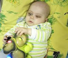
A Case of Acute Leukemia in a Baby
Авторы: Bogadelnikov I.V., Usova S.V., Dyabina T.A., Chvetko S.T., Vyaltseva Y.V., Crimean Medical University named.after S.I.Georgievsky, Simferopol, Ukraine, Regional Childrens Hospital, Simferopol, Ukraine.
Рубрики: Педиатрия/Неонатология, Онкология
Разделы: Справочник специалиста
Версия для печати
В детской инфекционной больнице находилась больная с кишечной инфекцией, у которой в дальнейшем развилась клиника сепсиса с резкой анемией и лейкемоидной реакцией. Установление диагноза врожденного лейкоза представляло объективные трудности.
У дитячiй iнфекцiйнiй лiкарнi перебувала хвора з кишковою iнфекцiєю, у якої надалі розвинулася нетипова клiнiка сепсису з рiзкою анемiєю та лейкемоїдною реакцiєю. Встановлення дiагнозу вродженого лейкозу становило об’єктивнi труднощi.
Female patient with intestinal infection, which later had a clinical picture of sepsis with severe anemia and leukemoid reaction, stayed in children’s infectious diseases hospital. The diagnosis of congenital leukemia had objective difficulties.
острый лейкоз, ребенок раннего возраста.
гострий лейкоз, дитина першого року життя.
acute leukemia, infant.
A 2.5 months-old girl admitted to a children’s infectious diseases hospital was diagnosed with an acute intestinal infection: acute enterocolitis. The child was suffering from: a fever 38.6 - 39.0°C, greenish mucous diarrhea, and loss of appetite.
The child was born on time after her mother’s third pregnancy that was not accompanied by any pathologies. There had been no history of any disease up to that age. After the birth, the child had a soft occipital swelling under the skin up to 1 cm deep, which soon disappeared leaving no traces.
The girl got sick acutely with body temperature rising up to 39°C. During the medical examination, her condition was characterized as moderately severe; the patient was fully conscious. The skin color was normal. Turgor and elasticity of tissues were preserved. Meningeal symptoms were absent. The prefontanel was 2 x 2 cm, normotonic. The peripheral lymph nodes were up to 0.3 cm, soft, movable. The tongue was moist, mildly coated. In the lungs, there was puerile respiration, no dyspnea. The cardiac tones were clear, rhythmic. The stomach was soft, no pain in the abdominal area reported. The liver was palpable for 2 cm under the costal arch, the spleen was not palpable. The patient’s stool was watery up to 4 - 5 times a day, bright yellow, green with mucous, undigested. The urine output was sufficient.
In spite of an antibiotic therapy going on for 9 days, the child continued having fever and diarrhea syndrome.
CBC showed marked decrease of Hb – 65 g/l and RBC – 2,3х1012, colour index 0,85. WBC 3,8х109, ESR 26 mm/h, stabs - 4%, segs – 20%, lymph – 67%, mn – 6%., PLT 196, hypochromia ++, anisocytosis ++, poikilocytosis ++ .
The blood group: АВ (4), Rh – factor (+) positive
Culture of breast milk: growth of S. epidermidis,
Stool test: Le 25-30 in the visual range, neutral fat – moderate amount, mucous – ++.
Stool culture sample for intestinal group of bacteria: negative
Crude protein: 46 g / l, albumin 28.5 g / L, urea 3.2 mmol / L, creatinine 0.068 mmol / L;
General bilirubin: 11 mmol / l, direct 4 mmol / l, indirect 7 mmol / L, AST - 0.34 mmol / L, ALT - 0.42 mmol / L, thymol test 2.6 units.
Coagulation: clotting time Lee-White - 11min 10 sec, prothrombin index 64%, fibrinogen A-1.75 g / l, the recalcification time 2min 35 sec.
The child was transfused with packed red blood cells -50 ml, IV and antibiotic therapy. The hemoglobin level was increased up to 105 g / l.
Later, the patient's condition remained severe due to intoxication syndrome, hyperthermia, diarrhea syndrome and (again) increasing anemia.
ELISA for TORCH infections showed Ig G of CMV, HSV и Toxo. Ig М to these pathogens were negative.
After the treatment, the child's body temperature was normal for 4 days, but the stool remained unstable. The mother and the child voluntarily left the hospital.
For the second time the child was admitted to the hospital 4 days later, diagnosed with an enterovirus infection, leukemoid reaction lymphocytic type, 3 degree anemia.
At the time of hospitalization, the child had fever (up to 39.5 C), watery stool, was vomiting, restless. On Rx of the chest, signs of a destructive right-sided medium-lobar pneumonia were detected.
An ultrasound of the abdomen detected liver abscess.
During the next 2 weeks, the child's condition deteriorated; signs of respiratory failure were observed; the hemoglobin level severely decreased again. At an examination, multiple soft-elastic consistency swellings were again detected on the scalp, not welded to the bones of the skull, immovable. At Rx of the skull bones, round bone defects were detected.
Subsequent bone marrow aspiration was done from the iliac bone: blasts - 36%, promyelocytes - absent, myelocytes - 1.5%, lymphocytes 3%, stab - 4%, segs - 2%, monocytes -10%, eosinophils - absent., normocytes basophilic - 2.5%, normocytes polychromatic - 17%, oxyphilic - 24%.
The bone marrow was moderately rich with cellular elements, cells of erythropoiesis - 43.5%. megacaryocytes: single.
With symptoms of multiple organ failure, a 2-3 degree coma developed, and the child died on the second day after being readmitted to the hospital.
Postmortem diagnosis: acute lymphoblastic leukemia.
Complication: bilateral confluent lobular pneumonia. Enterocolitis. DIC (fibrin clots and red blood cell sludge in the lumen of the capillaries, focal nephronecrosis, bleeding in parenchymatous organs)
Respiratory distress syndrome. Brain edema and swelling of the brain and the meningeals. Encephalopathy. Venous engorgement and parenchymatous degeneration of internal organs. Focal fatty liver. Accidental thymus conversion- 4 phase.
1. Врожденный лейкоз у детей / В.П. Булатов, Л.К. Фазлеева, И.Н. Черезова и др. // Казанский медицинский журнал. — 2001. — № 5. — С. 353356.
2. Вайнер М.А., Кейро М.С. Секреты детской онкологии и гематологии / М.А. Вайнер, М.С. Кейро. — М.: Бином, Невский диалект, 2008. — 272 с.
3. Дроздов А.А. Заболевания крови: полный справочник: Полная классификация. Механизмы развития. Самые современные методы диагностики и лечения / А.А. Дроздов, М.В. Дрождова. — М.: ЭКСМО, 2008. — 607 с.
4. Коленкова Г.В. Маркеры острого лейкоза в диагнозе заболевания у детей / Г.В. Коленкова // Гематология и трансфузиология. — 2002. — № 2. — С. 2835.
5. Кузьмина Л.А. Гематология детского возраста. — М.: МЕДэкспрессинформ, 2001. — 399 с.
6. Современная лабораторная диагностика онкологических заболеваний у детей в Украине / Д.Ф. Глузман, Л.М. Скляренко, В.А. Надгорная и др. // Онкология. — 2009. — Т. 11, № 2. — С. 139143.
7. Цитомегаловирусная инфекция у ребенка, больного острым лейкозом / Ерина Т.А., Варфаломеева С., Тимаков А.М., Добреньков К.В. // Педиатрия. — 1998. — № 1. — С. 100102.